

 PDF
PDF ePub
ePub Citation
 Print
Print


Abstract
Objectives
Abnormalities in various subcortical regions have been reported in previous structural neuroimaging studies for schizophrenia. To understand the subcortical abnormalities as a whole, all subcortical regions should be explored in each subject unlike most previous studies. Here, we explored major subcortical structures using volume measurement and shape analysis for schizophrenic patients (SZ), their unaffected siblings (Sib) and healthy controls without affected sibling (HC).
Methods
Structural magnetic resonance images were acquired from 24 SZ, 24 Sib and 19 HC. Both segmentation and shape analysis for subcortical structures was performed using FMRIB Integrated Registration and Segmentation Tool integrated within the FSL software. The group comparison of subcortical volumes was performed with multivariate analysis of variance (MANOVA).
Results
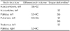
In SZ group, shape deformities were observed in the left nucleus caudates, left thalamus, left putamen and bilateral pallidus were increased compared with HC group. In Sib group, shape deformities were observed in the left pallidus, left putamen and left putamen was decreased compared with HC group. In Sib group, left nucleus accumbens was increased compared with SZ group.
Figures and Tables
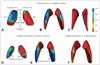 | Fig. 1Significant shape differences between different subject groups. For the SZ versus HC comparison, there are green-to-blue shading which denotes regions of inward deformation compared with healthy controls without affected sibling in the anterolateral part of the left thalamus (A), the anterolateral part of the left putamen (B) and the posteromedial part of the left nucleus caudates (C). For the Sib versus HC comparison, unaffected siblings showed deformity of the left globus pallidus (D), the left putamen (E) compared with healthy controls without affected sibling. Figures represent inward deformation in posterolateral region, outward deformation in posteromedial region of both structures. For the SZ versus Sib comparison, unaffected siblings showed outward deformation of left putamen (F) compared with schizophrenic patients. But there were no significant differences between schizophrenic patients and unaffected siblings after multiple comparison correction. SZ : Schizophrenic patients, Sib : Unaffected siblings, HC : Healthy controls without affected sibling.
|
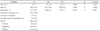
Table 1
Demographic data and clinical characteristics of schizophrenic patients (SZ), unaffected siblings (Sib), healthy controls without affected sibling (HC)
![]()
Table 2
Brain volume measurements of schizophrenic patients (SZ), unaffected siblings (Sib), healthy controls without affected sibling (HC)
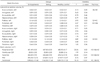
Data given as mean±SD, Volumes measured in cm3. A MANOVA was applied for estimating between group differences (p<0.05), Relative volume (%) represents absolute volume/Brain volume×100. CSF : Cerebrospinal fluid space, GM : Gray matter, WM : White matter, MANOVA : Multivariate analysis of variance, SD : Standard deviation
![]()
References
1. Ingvar DH, Franzén G. Abnormalities of cerebral blood flow distribution in patients with chronic schizophrenia. Acta Psychiatr Scand. 1974. 50:425–462.
2. Wright IC, Rabe-Hesketh S, Woodruff PW, David AS, Murray RM, Bullmore ET. Meta-analysis of regional brain volumes in schizophrenia. Am J Psychiatry. 2000. 157:16–25.
3. Honea R, Crow TJ, Passingham D, Mackay CE. Regional deficits in brain volume in schizophrenia: a meta-analysis of voxel-based morphometry studies. Am J Psychiatry. 2005. 162:2233–2245.
4. Witthaus H, Mendes U, Brüne M, Ozgürdal S, Bohner G, Gudlowski Y, et al. Hippocampal subdivision and amygdalar volumes in patients in an at-risk mental state for schizophrenia. J Psychiatry Neurosci. 2010. 35:33–40.
5. Harms MP, Wang L, Mamah D, Barch DM, Thompson PA, Csernansky JG. Thalamic shape abnormalities in individuals with schizophrenia and their nonpsychotic siblings. J Neurosci. 2007. 27:13835–13842.
6. Konick LC, Friedman L. Meta-analysis of thalamic size in schizophrenia. Biol Psychiatry. 2001. 49:28–38.
7. Staal WG, Hulshoff Pol HE, Schnack H, van der Schot AC, Kahn RS. Partial volume decrease of the thalamus in relatives of patients with schizophrenia. Am J Psychiatry. 1998. 155:1784–1786.
8. Andreasen NC. The role of the thalamus in schizophrenia. Can J Psychiatry. 1997. 42:27–33.
9. Weinberger DR. On localizing schizophrenic neuropathology. Schizophr Bull. 1997. 23:537–540.
10. Portas CM, Goldstein JM, Shenton ME, Hokama HH, Wible CG, Fischer I, et al. Volumetric evaluation of the thalamus in schizophrenic male patients using magnetic resonance imaging. Biol Psychiatry. 1998. 43:649–659.
11. Lipska BK, Jaskiw GE, Weinberger DR. Postpubertal emergence of hyperresponsiveness to stress and to amphetamine after neonatal excitotoxic hippocampal damage: a potential animal model of schizophrenia. Neuropsychopharmacology. 1993. 9:67–75.
12. Wright IC, Ellison ZR, Sharma T, Friston KJ, Murray RM, McGuire PK. Mapping of grey matter changes in schizophrenia. Schizophr Res. 1999. 35:1–14.
13. Wolkin A, Rusinek H, Vaid G, Arena L, Lafargue T, Sanfilipo M, et al. Structural magnetic resonance image averaging in schizophrenia. Am J Psychiatry. 1998. 155:1064–1073.
14. Bouix S, Pruessner JC, Louis Collins D, Siddiqi K. Hippocampal shape analysis using medial surfaces. Neuroimage. 2005. 25:1077–1089.
15. Mamah D, Harms MP, Wang L, Barch D, Thompson P, Kim J, et al. Basal ganglia shape abnormalities in the unaffected siblings of schizophrenia patients. Biol Psychiatry. 2008. 64:111–120.
16. Correa M, Carlson BB, Wisniecki A, Salamone JD. Nucleus accumbens dopamine and work requirements on interval schedules. Behav Brain Res. 2002. 137:179–187.
17. Grace AA. Gating of information flow within the limbic system and the pathophysiology of schizophrenia. Brain Res Brain Res Rev. 2000. 31:330–341.
18. Early TS, Posner MI, Reiman EM, Raichle ME. Hyperactivity of the left striato-pallidal projection. Part I: Lower level theory. Psychiatr Dev. 1989. 7:85–108.
19. Hokama H, Shenton ME, Nestor PG, Kikinis R, Levitt JJ, Metcalf D, et al. Caudate, putamen, and globus pallidus volume in schizophrenia: a quantitative MRI study. Psychiatry Res. 1995. 61:209–229.
20. Heckers S. Neuropathology of schizophrenia: cortex, thalamus, basal ganglia, and neurotransmitter-specific projection systems. Schizophr Bull. 1997. 23:403–421.
21. Chakos MH, Lieberman JA, Bilder RM, Borenstein M, Lerner G, Bogerts B, et al. Increase in caudate nuclei volumes of first-episode schizophrenic patients taking antipsychotic drugs. Am J Psychiatry. 1994. 151:1430–1436.
22. Lawrie SM, Abukmeil SS. Brain abnormality in schizophrenia. A systematic and quantitative review of volumetric magnetic resonance imaging studies. Br J Psychiatry. 1998. 172:110–120.
23. Velakoulis D, Wood SJ, Wong MT, McGorry PD, Yung A, Phillips L, et al. Hippocampal and amygdala volumes according to psychosis stage and diagnosis: a magnetic resonance imaging study of chronic schizophrenia, first-episode psychosis, and ultra-high-risk individuals. Arch Gen Psychiatry. 2006. 63:139–149.
24. Hwang J, Lyoo IK, Dager SR, Friedman SD, Oh JS, Lee JY, et al. Basal ganglia shape alterations in bipolar disorder. Am J Psychiatry. 2006. 163:276–285.
25. Byne W, Buchsbaum MS, Kemether E, Hazlett EA, Shinwari A, Mitropoulou V, et al. Magnetic resonance imaging of the thalamic mediodorsal nucleus and pulvinar in schizophrenia and schizotypal personality disorder. Arch Gen Psychiatry. 2001. 58:133–140.
26. Lehéricy S, Ducros M, Van de Moortele PF, Francois C, Thivard L, Poupon C, et al. Diffusion tensor fiber tracking shows distinct corticostriatal circuits in humans. Ann Neurol. 2004. 55:522–529.
27. Kandel ER, Schwartz JH, Jessell TM. Principles of neural science. 2000. 4th ed. New York: McGraw-Hill.
28. Yazici AH, Demir B, Yazici KM, Göğüş A. Neurological soft signs in schizophrenic patients and their nonpsychotic siblings. Schizophr Res. 2002. 58:241–246.
29. Krauzlis RJ. Recasting the smooth pursuit eye movement system. J Neurophysiol. 2004. 91:591–603.
30. Abel LA, Levin S, Holzman PS. Abnormalities of smooth pursuit and saccadic control in schizophrenia and affective disorders. Vision Res. 1992. 32:1009–1014.
31. Boos HB, Aleman A, Cahn W, Hulshoff Pol H, Kahn RS. Brain volumes in relatives of patients with schizophrenia: a meta-analysis. Arch Gen Psychiatry. 2007. 64:297–304.
32. Scherk H, Falkai P. Effects of antipsychotics on brain structure. Curr Opin Psychiatry. 2006. 19:145–150.
33. Margolese HC, Malchy L, Negrete JC, Tempier R, Gill K. Drug and alcohol use among patients with schizophrenia and related psychoses: levels and consequences. Schizophr Res. 2004. 67:157–166.
 XML Download