

 PDF
PDF ePub
ePub Citation
 Print
Print


INTRODUCTION
Esophageal perforation is one of the most serious complications of atrial fibrillation (AF) ablation, with an incidence ranging from 0.016–0.04%.1)2)3) Left atrial-esophageal fistula frequently leads to systemic embolization or death and usually requires surgical management.4)5) However, it has been reported that pericardial-esophageal fistula can be managed non-surgically.6)7)8)9) We report a case of pericardial-esophageal fistula complicating AF ablation that was successfully managed with a conservative approach.
CASE
A 40-year-old male patient was referred to our hospital for paroxysmal AF with palpitation. The patient had been taking immunosuppressive medication for rheumatoid arthritis for 8 years. Prior to AF ablation, the patient was taking prednisolone (20 mg/day) and hydroxychloroquine (300 mg/day). Because anti-arrhythmic therapy with flecainide or propanone failed to suppress symptomatic paroxysmal AF recurrence, we performed radiofrequency catheter ablation. With the patient under general anesthesia with mechanical ventilation, ablation was performed using a 3-dimensional navigation system (CARTO 3 V3.2 EP Navigation System; Biosense Webster Ltd., Tirat Hacarmel, Israel) and a 7.5F steerable irrigation tip ablation catheter (ThermoCool® SF, Bi-Directional NAV Catheter; Biosense Webster Ltd., Diamond Bar, California, USA). Electrical isolation of the pulmonary vein (PV) from the left atrial body was performed via bi-antral and bi-carinal ablation. Esophageal temperature monitoring and intracardiac echocardiography were not available during ablation. Ablation power was set to 30 watts for the left atrial anterior wall and 25 watts for the posterior wall, with a temperature cutoff of 40°C. Lower and upper impedance cutoffs were 50 ohms and 250 ohms, respectively. Radiofrequency ablation was performed in AF. Although ablation was primarily guided by the changes in PV potentials, we tried to deliver radiofrequency energy for no more than 40 seconds (20–30 seconds for the posterior wall) at each ablation point in order to avoid excessive tissue ablation. The total procedure time was 7 hours and 10 minutes, and total left atrial ablation time was 2 hours and 6 minutes. Ablation times for the initial left- and right-side PV isolation were 42 and 36 minutes, respectively. Ablation time for the left side posterior wall was approximately 13 minutes.
During left-side posterior wall ablation, linear ablation was performed at the site of greatest proximity to the esophagus (Figure 1); however, ablation was not interrupted by increased temperature or impedance. Because PVs were repeatedly reconnected to the left atrium after isoproterenol infusion at infusion rates of 1–3 µg/min, additional ablations were performed for 48 minutes along the linear ablation lines to complete electrical PV isolation or inside the PVs to abolish dissociated PV potential. Finally, complete PV isolation was achieved, and the ablation procedure was finished without acute complications after cavotricuspid isthmus ablation.
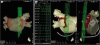
Figure 1
Radiofrequency ablation findings. (A, B) Electroanatomic mapping images of the left atrium acquired by the CARTO 3 Navigation System show that linear ablation at the posterior wall was performed in front of the esophagus during left-side antral ablation. (B) Intracardiac electrograms recorded by the ablation catheter (AB 1–2 and AB 3–4) during left-side posterior wall ablation show stable left atrial potentials that ranged from 0.8–1.4 mV. The tip of the ablation catheter tightly contacts the left atrial endocardium at a less than 2 mm distance to the esophagus.
AB = ablation catheter electrodes; CS = coronary sinus catheter electrodes.
Following AF ablation, the patient began complaining of persistent retrosternal pain, which required fentanyl-patch application and intermittent morphine injections (Figure 2). Pantoprazole (40 mg/day) was empirically administered intravenously to reduce the risk of esophageal ulcer development. Because symptomatic AF recurred 3 days after ablation, bisoprolol (2.5 mg/day), and flecainide (150 mg/day) were administered. The patient was discharged following 5 days of significant improvement in retrosternal pain. However, the patient visited the emergency room 9 days after ablation for recurring symptomatic AF. The patient complained of mild but persistent chest pain with paroxysmal palpitation, which persisted for 2–3 hours, recurring every day after hospital discharge despite adherence to anti-arrhythmic medications.
Eventually, 17 days following AF ablation, the patient visited the emergency room again for chest pain, which had become aggravated by a deep inspiration and coughing and required him to remain prone. However, the patient did not complain of odynophagia. A fever of 38°C with severe chills and leukocytosis (white blood cell count: 15,180/µL) were noted. Surface electrocardiogram showed a normal sinus rhythm of 78 bpm without significant changes in ST-segments or QRS voltages, and cardiac troponin-I and C-reactive protein levels were within normal reference ranges. There was no cardiomegaly or pleural effusion on the initial chest X-ray image (Figure 3A). Abnormal pericardial fluid collection was not detected by bedside echocardiography.
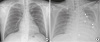
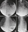
Figure 3
Chest X-ray findings. (A) Initial chest X-ray image in posteroanterior projection does not show cardiomegaly or pleural effusion. (B) Chest X-ray image acquired 2 days after esophageal perforation shows an air-line and bubbles within the cardiac silhouette (arrows) in anteroposterior projection.
Chest computed tomography (CT) performed in the emergency room showed small but clustered air bubbles in the pericardial space, suggestive of esophageal perforation (Figure 4A); however, neither the radiologists nor the cardiologist in the emergency room could identify the presence of air bubbles in the pericardial space. Because an esophageal injury was suspected, the patient was admitted to the sub-intensive care unit for close monitoring; nevertheless, no radiologic signs of esophageal injury were noted by the physicians, and the patient was erroneously permitted to take small sips of water to quench his thirst, which could have aggravated mediastinitis. Although broad-spectrum antibiotics were administered intravenously, within 2 days, the patient rapidly proceeded to sepsis that was complicated by multi-organ failure. A rapid increase in pericardial and pleural effusions with the appearance of large air bubbles within the pericardial space was noted on chest X-ray 2 days after admission (Figure 3B). Esophagography performed 3 days after admission confirmed leakage of contrast agent into the pericardial space, but not into the left atrium (Figure 5A and 5B). Because clinical signs of communication between the left atrium and the esophagus, such as neurologic abnormalities or hematemesis, were not observed for 3 days after admission, the possibility of left atrial-pericardial fistula formation was regarded as low. No air bubbles in the left atrium on the initial chest CT scan and contrast agent leakage into the left atrium on the initial esophagography also indirectly supported the conclusion of no left atrial-esophageal fistula. After discussing the case with thoracic surgeons, we opted to continue with conservative minimally invasive pericardial management and pleural drainage to avoid the complications associated with major surgery. Endoscopic intervention was considered as a secondary treatment option. Emergent pericardiostomy and chest tubing were performed to remove infected mediastinal fluids. During the pericardiostomy while the patient was under local anesthesia, we repeatedly checked his mental status and performed serial neurologic examinations to recognize a potential embolic stroke as soon as possible. Because a thoracoscope was not available at the time, the operating thoracic surgeon had to indirectly confirm via visual inspection of the drained pericardial fluid that there was no active bleeding from the left atrium. A pericardial drainage tube was gently inserted and placed in the pericardial space without further injury. Neurologic signs caused by systemic air embolism did not appear during pericardiostomy.
Figure 4
Chest CT scan findings. (A) An initial chest CT image shows clusters of small air bubbles (black arrow) in the pericardial space, which is highly suggestive of an esophageal perforation. However, there were no air bubbles within the left atrium. Left atrial posterior wall thickness was less than 2 mm, and intervening connective and fat tissues between the left atrial posterior and esophageal walls were virtually absent. (B) Chest CT image acquired 22 days after esophageal perforation shows large air bubbles (white arrow) remaining in the pericardial space. However, there were no further complications.
CT = computed tomography.
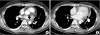
Figure 5
Esophagography findings. Initial esophagography shows contrast agent (arrows) leaking from the mid-esophagus into the pericardial space at the level of 9 cm above the esophagogastric junction (A, B). Follow-up esophagography shows no more contrast agent leakage into the pericardial space (C, D). Radiologic findings that reveal the presence of esophageal ulcer or stricture were not observed on the initial or follow-up esophagography images.
Immediately after starting drainage of pericardial and pleural effusion, fever was resolved, and the patient's general condition improved rapidly. Because there were no signs of left atrial-esophageal fistula for 3 days after admission, including massive gastrointestinal tract bleeding or systemic air embolism, and because the pericardial-esophageal fistula did not appear to be complicated by left atrial-esophageal fistula, we decided to continue conservative management based on very strict and prolonged fasting with fluid replacement, broad-spectrum antibiotic therapy, and continuous drainage of the infected pericardial and pleural effusions. The patient was even instructed to spit out saliva and not to ingest any food or water. Total parenteral nutrition was performed during the fasting period. Pericardial and pleural effusions were continuously removed via pericardial and bilateral chest tubing. The patient recovered from sepsis and multi-organ failure after 3 days of drainage. Follow-up esophagography performed 15 days after pericardiostomy showed no contrast agent leakage into the pericardial space (Figure 5C and 5D). Dietary intake was then restarted and increased gradually with close monitoring of the patient's symptoms, vital signs, chest X-ray, and laboratory findings. Follow-up chest CT performed 20 days after pericardiostomy showed improving mediastinitis (Figure 4B). The patient was discharged 28 days after pericardiostomy without further complications. The recurrence of symptomatic AF was suppressed by antiarrhythmic therapy at the time of hospital discharge. Administration of antiarrhythmic agents was stopped 3 months after ablation.
DISCUSSION
Esophageal perforation is a rare but severe complication following radiofrequency catheter ablation of AF. Prior worldwide surveys reported that the incidence of esophageal perforation-related complications after AF ablation ranged from 0.016% to 0.04%.1)2)3) However, incidences of esophageal perforation at small-volume centers might be underreported, and some cases might prove fatal before a confirmatory diagnosis.4) Therefore, the actual incidence of esophageal perforation might be higher than that reported in the literature. Furthermore, subclinical esophageal injury might be much more prevalent than clinically manifested esophageal perforation. A previous study in which endoscopic examination was performed in 28 patients who underwent AF ablation reported that approximately 50% of the patients experienced esophageal wall changes within 24 hours.10) Another study in which endoscopic ultrasonography was performed for 29 patients who underwent AF ablation reported that approximately 30% of the patients had morphologic changes of the periesophageal connective tissues and the left atrial posterior wall without esophageal mucosal erythema or ulcers.11) Therefore, all patients who undergo AF ablation should be regarded as having esophageal and periesophageal tissue injury with an associated risk of esophageal perforation.
The pathophysiologic mechanisms of pericardial-esophageal fistula formation are not well understood. Direct thermal injury, ischemic injury by thrombotic occlusion of end arterioles, chemical injury by digestive enzymes and gastric acid, or bacterial infection of the lumen are all regarded as causes of delayed esophageal fistula formation.12)13) Technical factors related to esophageal injury include the total amount of radiofrequency energy delivered to the ablation sites near the esophagus, use of large-tip or irrigation-tip catheters, vertical catheter orientation, and contact pressure.14) Tissue factors related to esophageal injury include atrial wall thickness and thickness and characteristics of connective tissue between the left atrial and esophageal walls.14) Ablation with the patient under general anesthesia, compared with conscious anesthesia, is also known to increase the risk for esophageal injury.15) In this case, ablation was performed using an irrigation-tip catheter under general anesthesia. During left-side antral isolation, the left atrial posterior wall was ablated for about 13 minutes at 25 watts, suggesting that the total radiofrequency energy delivered at the left atrial posterior wall was not excessive. However, the ablation line generated at the left atrial posterior wall was very close to the esophagus (Figure 1). The shortest distance between the endocardial ablation line and esophageal wall was less than 2 mm on CARTO images. Because ablation was performed with the patient under general anesthesia, catheter positioning and contact were very stable, and continuous ablation was performed using an irrigation-tip catheter without temperature interruptions or impedance cutoff. Additionally, the left atrial posterior wall thickness was less than 2 mm, and intervening connective and fat tissues between the left atrial posterior and esophageal walls were nearly absent (Figure 4A). Methylprednisolone taken for rheumatoid arthritis could have prevented the regeneration of esophageal tissue following thermal injuries.
Left atrial-esophageal fistula is usually fatal if complicated by systemic embolism or massive bleeding from the gastrointestinal tract.4)5) Although high fever or chest pain appearing 1–4 weeks after AF ablation are typically the first signs of left atrial-esophageal fistula formation, there are no specific symptoms, signs, or tests to predict the development of esophageal perforation. Clinical detection of a pericardial-esophageal fistula can be difficult because conspicuous signs, such as systemic embolization and gastrointestinal tract bleeding, are not present.6)7)8)9) Therefore, to recognize the condition and avoid delay of an appropriate diagnosis, clinical suspicion should increase if there are any chest symptoms or signs suggestive of extensive tissue injury. In this case, after AF ablation, the patient complained of persistent retrosternal pain for more than 9 days, which is suggestive of extensive tissue injury. However, the patient did not complain of odynophagia even after the esophageal perforation. The frequent recurrence of AF refractory to anti-arrhythmic therapy despite successful PV isolation, might be indicative of persistent inflammation with atrial tissue irrigation. This case indicates that unusually persistent and severe post-ablation chest pain and an unusually frequent recurrence of AF despite successful ablation should be considered as possible signs of extensive tissue injury with increased risk of esophageal injury. Although there were clinical manifestations suggestive of the presence of an esophageal injury, we failed to identify the air bubbles in the pericardial space because we were so focused on confirming air bubbles in the left atrium, which would have indicated the presence of left atrial-esophageal fistula. In prior case reports, pericardial-esophageal fistula presence was suspected by the presence of air, pericardial effusion, or contrast agent leakage into the pericardial space on initial chest CT scans.4)5)6)7) However, in our case, radiologic signs of esophageal injury were not present on the chest CT scan at the time of initial presentation except for small air bubbles in the pericardial space, which can only be recognized with careful inspection. Because these radiologic signs were not recognized, the patient was permitted small sips of water, which might have aggravated mediastinitis; esophagography was performed 2 days later. This case demonstrates that careful review of chest CT images by experienced radiologists and cardiologists is very important, and emergent esophagography should be performed when there is sufficient clinical suspicion to avoid a delay in diagnosis. Fortunately, when compared with left atrial-esophageal fistula, pericardial-esophageal fistula has a relatively benign course. Previous case studies reported that pericardial-esophageal fistula was successfully treated with supportive care or endoscopic intervention.6)7)8)9) In this case, pericardial-esophageal fistula was successfully healed by very strict fasting over 2 weeks with fluid and antibiotic therapy after drainage of inflammatory body fluids. Although a proton pump inhibitor was empirically administered after ablation due to the possibility of esophageal injury, esophageal perforation eventually developed. There is no clinical evidence that anti-ulcer medications, such as proton pump inhibitors or sucralfate, can reduce the incidence of esophageal perforation. Therefore, if esophageal perforation is suspected, strict fasting and broad-spectrum antibiotic therapy should be initiated as soon as possible, even before confirmation of esophageal perforation.
In summary, we report a case of pericardial-esophageal fistula complicating AF ablation that was successfully treated by conservative management. Pericardial-esophageal fistula is a very rare complication of AF ablation. If diagnosed early, it can be treated by conservative management, including pericardial drainage. Appropriate radiologic imaging studies under conditions of high clinical suspicion should be performed immediately to avoid a delay in diagnosis.
 XML Download