

 PDF
PDF ePub
ePub Citation
 Print
Print


Introduction
Radiofrequency catheter ablation (RFCA) as a treatment option for ventricular tachycardia (VT) is an important non-pharmacological alternative or adjunct to antiarrhythmic agents.1) Unfortunately, many cases of VT with or without underlying structural abnormalities are considered ineligible for mapping and ablation largely due to hemodynamic instability.2) Under these circumstances, hemodynamic support during the procedure enables detailed activation and entrainment mapping to guide successful ablation.3) Several previous studies demonstrated the use of percutaneous left ventricular (LV) assist devices (pLVAD), such as extracorporeal membrane oxygenation, Impella or TandemHeart system-supported RFCA for hemodynamically unstable VT.3)4)5) Intravenous (IV) inotropic agents are adjunctive in these cases, but the safety and efficacy of IV dopamine alone for hemodynamic support remains unknown. We presented seven patients with successful RFCA under support with IV dopamine alone for poorly tolerated idiopathic VT.
Subjects and Methods
Study population and definition
A total of 86 patients underwent de novo RFCA for idiopathic VT between 2010 and 2015 at our institution. All cases were reviewed retrospectively, and 7 (8.1%) consecutive patients in whom IV dopamine was applied for hemodynamic support during RFCA were enrolled in the study. Hemodynamically unstable VT was defined as a drop in mean blood pressure (BP) <70 mmHg and/or requiring direct current (DC) cardioversion due to induced VT during the procedure. Electrical storm was defined as ≥3 separate episodes of ventricular arrhythmias within a 24-hour time period. Informed consent was obtained from all patients, and the study was approved by the Institutional Review Board of Korea University Medical Center.
Electrophysiologic study, mapping and ablation
One patient was prescribed an antiarrhythmic drug (AAD), amiodarone, which was discontinued 4 weeks before the procedure. All procedures were performed under deep sedation with IV propofol, and oropharyngeal airways were maintained. Blood pressure was closely monitored via a transfemoral arterial approach. Programmed electrical stimulations (PES), including rate-incremental pacing and up to 3 extrastimuli pacing at the right ventricle (RV) and/or LV were applied for VT induction with or without isoproterenol. VT was induced in all cases. Non-clinical VT was also induced in one case, but only clinical VT was targeted for ablation.
Activation mapping was attempted in all cases. For hemodynamic instability due to VT, IV dopamine was administered at an initial dose of mean 6.2±1.3 mcg/kg/min, and then the dosage was adjusted based on changes in mean BP to maintain at least 70 mmHg during VT. As long as VT was sustained stably, it was not prematurely terminated with PES or cardioversion, and mapping was continued. Entrainment (2 cases, 28.6%) and/or pace mapping (4 cases, 57.1%) was added to supplement substrate or activation mapping. Intracardiac echocardiography (AcuNav, Siemens Healthcare, Issaquah, WA, USA) and a three-dimensional electroanatomic mapping system (NavX system; St. Jude Medical Inc., St. Paul, MN, USA and Carto-3 system; Biosense Webster, Inc., Diamond Bar, CA, USA) were applied in 4 (57.1%) and 3 cases (42.9%), respectively. In 2 cases (28.6%), epicardial mapping was performed via a percutaneous subxiphoid approach.
Radiofrequency energy was delivered via a 4 mm open irrigated tip catheter (Coolflex; St. Jude Medical Inc., St. Paul, MN, USA) in 2 cases (28.6%) or a non-irrigated catheter (Blazer II, Boston Scientific Inc., Natick, MA, USA) in remaining 4 cases (57.1%). Acute procedural success was defined as the termination and the lack of inducibility of clinical VT with or without isoproterenol after ablation.
Follow-up
All patients were followed 2 weeks after the procedure and subsequently, every 3-6 month for at least one year. At every visit, any symptoms, surface electrocardiogram (ECG) and 24hr Holter monitoring were checked to confirm the absence of recurrence. An implantable cardioverter-defibrillator (ICD) implanted in one patient was interrogated at each follow-up visit. AAD was not prescribed after the procedure.
Statistical analysis
Continuous variables were analyzed for normality using Kolmogorov-Smirnov test. Normally distributed data were represented as mean±standard deviation, and the others as the median (25 percentile, 75 percentile). Categorical variables were expressed as counts and percentages. Statistical analyses were performed using SPSS Statistics version 21.0 (Statistical Package for the Social Sciences; SPSS Inc., Chicago, IL, USA).
Results
Baseline clinical characteristics
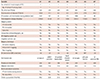
The baseline clinical characteristics were summarized in Table 1. All patients were men with a mean age of 50.7±5.3 years. Clinical VT was sustained requiring cardioversion in 4 patients (57.1%), and of these, 2 patients experienced VT storm. Transthoracic echocardiography showed normal biventricular function and absence of any structural abnormalities. Coronary angiography in 4 patients, cardiac magnetic resonance or computed tomography imaging in 2 patients and exercise ECG in 1 patient indicated no evidence of ischemia. ICD was implanted in one patient for secondary prevention for a prior attack of hemodynamically unstable VT. Surface ECG of clinical VT indicated that all were monomorphic, with right bundle branch block (RBBB) pattern in 4 cases and inferior QRS axis in 5 cases. At presentation, 3 patients (42.9%) were on beta-blockers, and one patient was on amiodarone.
Electrophysiologic and ablation characteristics
The summary of procedural characteristics of each patient was shown in Table 2. Non-clinical VT was induced in one patient, which was hemodynamically compromised and terminated by cardioversion. All remaining induced VTs were clinical and targeted for mapping and ablation. The average tachycardia cycle length was 281.7±13.0 ms. The mean total duration of induced VT was 35.9±15.3 minutes under dopamine support. VT was originated from LV in 6 patients (LV septum, aortic cusp, LV summit, LV apex, anterolateral papillary muscle, and left posterior fascicle) and RV outflow tract in 1 patient. Successful RFCA guided by activation mapping with mean earliest activation time before QRS onset of -38.7±7.2 ms; except for one fascicular origin VT that was successfully eliminated by targeting a Purkinje potential during VT was achieved in all patients (6 endocardial and 1 epicardial ablation); and non-inducibility was also achieved after ablation (Table 2). The median duration of ongoing VT until final termination was 2.8 (0.7, 21.4) minutes. The mean procedure and ablation times were 215.2±25.4 and 29.2±9.9 minutes, respectively.
Effects of intravenous dopamine administration on blood pressure
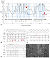
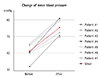
Premature VT termination with external cardioversion was required in 2 patients (28.6%) before dopamine infusion. These patients needed additional cardioversion even after dopamine infusion. Because one patient showed atrial fibrillation during VT, which was terminated by DC cardioversion, as shown in Fig. 1A-F. In the other patient, BP was unstable within the first several minutes immediately following dopamine infusion, but stabilized enabling activation mapping after the dopamine dosages were increased. The mean initial and the mean peak dosages were 6.17±1.30 mcg/kg/min and 14.86±3.83 mcg/kg/min, respectively. The average dose of dopamine in all patients was 1266.1±389.6 mcg/kg. The average mean BP during VT without dopamine was 52.3±4.1 mmHg and rose to 82.6±3.8 mmHg (Δ28.8±3.2 mmHg) after IV dopamine infusion (Fig. 2). No other tachyarrhythmias occurred, except one case of induced non-clinical VT, and increase in heart rate during sinus rhythm was not remarkable during dopamine infusion.
Clinical outcomes
After successful RFCA, no acute events such as cardiac ischemia, hypoxic brain damage, acute kidney injury or cognitive dysfunction occurred in any of patients. VT or premature ventricular contraction recurrence was absent in all patients without AAD, during a mean follow-up period of 23.0±6.1 months.
Discussion
We reported seven patients with hemodynamically unstable idiopathic VT in which RFCA was performed successfully with IV dopamine support. Improved hemodynamic stability provided by dopamine permitted sufficiently prolonged VT duration for detailed activation and/or entrainment mapping, leading to procedural success.
Mapping strategies in poorly tolerated VT
Conventional technique such as activation and entrainment mapping or substrate-based mapping technique is selected based on the etiology and hemodynamic tolerance of VT.6) Activation and entrainment mapping is difficult, since induced clinical VT is often poorly tolerated. Recent studies demonstrated a substrate-based ablation strategy targeting late potentials, conductive channels, linear lesions, or local abnormal ventricular activities during sinus rhythm for hemodynamically unstable VT.7)8)9) However, different characteristics of substrates should be considered based on VT etiology.
A substrate-based ablation strategy is less effective in idiopathic VT, as compared to other premature ventricular contraction VT, due to a paucity of arrhythmia substrates in most cases.10)11)12) Pace mapping can be applied if VT is scarcely inducible or intolerable, but it also has some limitations, i.e., variability in the paced QRS or possible reproducibility of similar QRS morphology despite different origins due to close anatomical relationship.11)13)14) In this respect, activation and entrainment mapping to corroborate putative ablation sites is necessary in idiopathic VT. Furthermore, intolerance is reportedly an independent predictor of recurrence after ablation.15) Therefore, hemodynamic support is warranted to enable proper mapping and achieve better outcomes of RFCA for hemodynamically unstable idiopathic VT.
Selection of strategy for hemodynamic support during unstable VT ablation
Hemodynamic support during RFCA for hemodynamically unstable VT permits prolonged mapping and reduces the detrimental impact on end-organ function related to systemic malperfusion.11) Recently published data demonstrated that mechanical support with pLVAD allows for a longer duration of sustained VT, more adequate activation and entrainment mapping and more frequent VT termination during energy delivery.4)16)17) The efficacy of pLVAD is noticeable despite the lack of prospective randomized trials and the ongoing controversy over the rate of non-inducibility as a procedural endpoint or long-term freedom from VT.11)18) However, pLVAD is not always available in a VT ablation-capable laboratory. Time, cost and additional complications related to its implantation and removal are also problematic.19)
The role of dopamine on ablation of intolerable VT
Dopamine is inexpensive, familiar and readily available in the electrophysiologic (EP) laboratory. In our cases, IV dopamine infusion as the sole hemodynamic supporter was sufficient for conventional mapping and ablation for VT. Three patients (42.9%) were recommended to undergo ICD implantation rather than RFCA due to poorly tolerated VT requiring frequent premature termination during EP study before referral to our institute. Of these, only one patient received an ICD, but the other two refused. They have been free from VT after successful dopamine-supported ablation, and ICD in one patient went off neither antitachycardia nor shock therapy thereafter.
Dopamine has a dose-dependent proarrhythmic effect via increasing automaticity or a biphasic effect on action potential duration.20) In our study, fatal arrhythmia rarely occurred, which might be due to relatively low dosage of dopamine and structurally normal heart. In this study, only dopamine was tried, but other inotropics such as dobutamine, phenylephrine, epinephrine, norepinephrine, or milinone might be also considered. A previous study reported the use of dobutamine and phenylephrine as well as dopamine for hemodynamic support, even though they were subsidiary to pLVAD.3) However, caution is required due to the proarrhythmic effects of these drugs.
Who is a good candidate for dopamine-supported VT ablation?
Since this was not a comparative trial, the relevant characteristics of patients who potentially benefited from dopamine support were not determined. Patients with idiopathic VT might be better candidates for dopamine use than those with structural heart disease associated VT, as they are relatively less vulnerable to hemodynamic instability with aggravation of ischemia during sustained VT. However, there is limited data regarding which type of clinical VT would become hemodynamically unstable during an EP study. Further studies are warranted to investigate whether VT etiology, LV systolic dysfunction, or history of DC cardioversion prior to the procedure is useful to identify good candidates for dopamine support. Additional studies may also determine whether patients with hemodynamically unstable VT can be mapped and ablated under dopamine support alone or whether more aggressive preparation such as pLVAD are needed.
Limitations
First, this was a retrospective single center analysis with a limited number of patients. Second, a possible negative inotropic effect of propofol that might affect hemodynamic stability during the procedure was not considered. In 2 patients, BP decreased significantly just after initiating sedation, however, it was stabilized after the dosage was reduced as protocol and compromised state did not occur thereafter unless VT was induced in both patients. Third, other measures for hemodynamic stability such as cerebral oximetry were not taken during the procedure. However systemic oxygen saturation and hepatic performance or renal function were within normal limits after the procedure, which suggested no deteriorating effects of VT with low pressure. Fourth, since this was a single-arm study, there was no data comparing the efficacy of RFCA with or without dopamine infusion. Despite these limitations, dopamine prevents early cessation of procedure without attempt of ablation due to hemodynamic instability.
In conclusion, IV dopamine provided hemodynamic support during ablation of idiopathic VT with hemodynamic instability and enabled detailed mapping leading to successful ablation.

 XML Download