

 PDF
PDF ePub
ePub Citation
 Print
Print


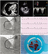
A 78-year-old man visited the emergency department with dyspnea of New York Heart Association Class III for 2 months. His past medical history comprised hypertension and old tuberculosis. He had no history suggesting coagulopathy, recurrent history of thromboembolism, or arrhythmia such as atrial fibrillation. He worked as a guard who spent every other night in the sitting position. The laboratory data were as follows: white blood cell count, 7910/mm3 (61.2% neutrophils, 27.4% lymphocytes, and 8.1% monocytes); hematocrit, 41.2%; platelets, 243000/mm3; bleeding time, 1 min; clotting time, 9 min; fibrinogen, 457 mg/dL (normal, 200-400 mg/dL); D-dimer, 7.1 µg/mL (normal, <0.5 µg/mL); factor V, 64% (normal, 60-140%); protein C antigen, 44% (normal, 72-160%); protein S antigen, 69% (normal, 60-150%); von Willebrand factor, 293.5%; and N-terminal pro B-type natriuretic peptide, 1660 pg/mL. Chest computed tomography (CT) showed thromboembolism of the right main pulmonary artery and both segmental pulmonary arteries (Fig. 1A). His echocardiography showed right ventricular (RV) dysfunction with hypokinesia in the enlarged RV and elevated RV systolic pressure (tricuspid regurgitation peak velocity: 3.98 m/s) and a D-shaped left ventricle (LV) suggesting pulmonary thromboembolism (PTE) (Fig. 1B, C). There was no evidence of other congenital abnormalities including persistent left superior vena cava on CT or echocardiography. Lower extremity Doppler ultrasonography also showed deep vein thrombosis of the muscular branches with dilation and obstruction. On the 8th hospital day, during anticoagulation treatment, he complained of chest pain and electrocardiography showed ST segment elevation in leads II, III, and aVF with reciprocal changes (Fig. 1D). Emergent coronary angiography revealed ball-like thrombotic total occlusion in the distal right coronary artery (RCA) (Fig. 1E). After 3 sessions of thrombus aspiration, massive red thrombi were obtained with TIMI 3 flow (Fig. 1F, G). There was no evidence of plaque rupture on intravascular ultrasound. The procedure was successfully completed without balloon angioplasty or stent implantation. We recommended interventional or surgical embolectomy of pulmonary arteries but the patient refused further interventions. After anticoagulation, his dyspnea improved. Follow-up chest CT showed slightly decreased but still persisting PTE in both pulmonary arteries. Follow up echocardiography showed slightly decreased RV diastolic area (from 30.82 to 26.42 mm2), decreased RV systolic pressure (from 75.29 to 59 mmHg) with resolution of the D-shaped LV, and it demonstrated no intracardiac thrombus and normal left atrial appendage mechanical function. We performed agitated saline contrast study to evaluate the presence of paradoxical embolic route such as a patent foramen ovale (PFO), and we demonstrated that agitated saline was seen in the left atrium within 3 beats during Valsalva maneuver (Supplementary Video in the online-only Data Supplement).
Myocardial infarction due to paradoxical embolism of a thrombus in the venous system is a rare phenomenon. In this case, a massive thrombus was detected in the RCA, which might be paradoxical thromboembolism via a PFO. Little evidence exists to guide the decision to close PFOs in such a situation, although small-sized studies have reported that PFO might be associated with adverse outcome in patients with PTE.
 XML Download