

 PDF
PDF ePub
ePub Citation
 Print
Print


Introduction
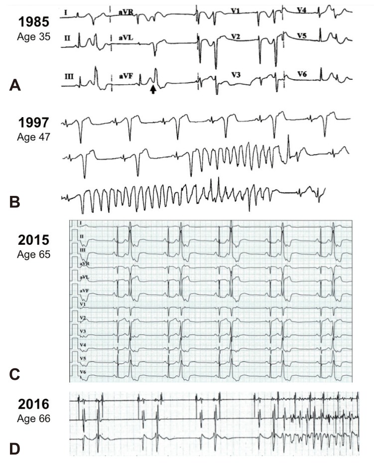
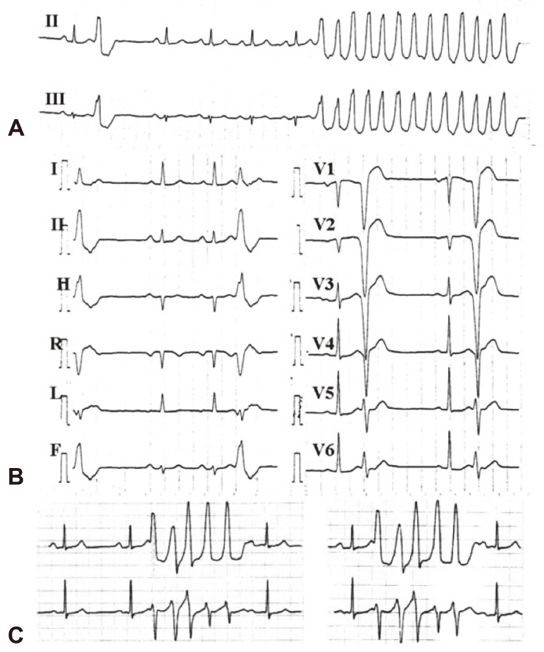
Ventricular extrasystole originating from the right ventricular outflow tract (RVOT) or the left ventricular outflow tract (LVOT) is the most commonly encountered ventricular arrhythmia in ostensibly healthy individuals with no evidence of heart disease.1)2) For example, among otherwise healthy athletes with ventricular arrhythmia, the arrhythmia site of origin is the ventricular outflow tract in roughly 70%.3) These ventricular arrhythmias have a characteristic electrocardiographic morphology, with a left bundle branch block pattern and tall R-waves in the inferior leads of either the normal or right axis (Fig. 1A, C, 2B).4) In fact, this QRS morphology is so distinctive that it is common practice to accept the diagnosis of “idiopathic benign ventricular arrhythmia from the outflow tract” based on this unique morphology when the electrocardiogram (ECG) during sinus rhythm and the echocardiogram are normal, sometimes negating the need for invasive tests.1)5)
A small proportion of patients with an unusually high burden of ventricular extrasystole can developing a reversible deterioration of left ventricular function as manifestation of “tachycardia-induced cardiomyopathy.”6) Still, the vast majority of patients with idiopathic outflow tract arrhythmias have an excellent long-term prognosis. Even if the outflow ventricular extrasystole ultimately triggers sustained ventricular arrhythmia, the resulting ventricular tachycardia (VT) will be a monomorphic VT originating from the outflow tract (Fig. 2A), which is a scenario that is known to be hemodynamically well tolerated.1)5) Thus, the idiopathic ventricular arrhythmias originating from the outflow tract are universally considered benign.1)2)4)7)
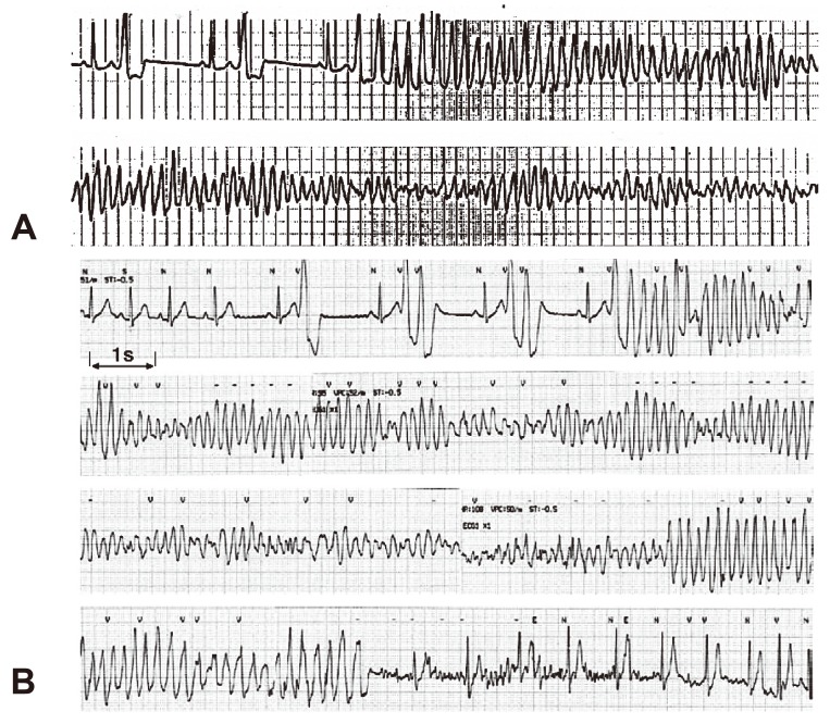
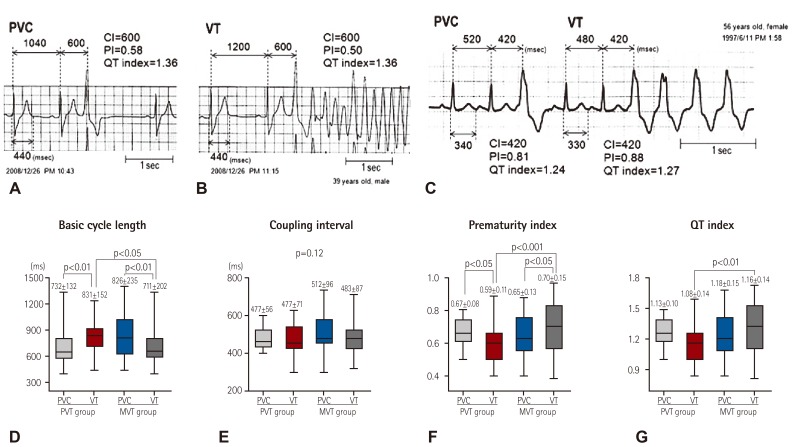
In 2005, we described three patients with typical idiopathic RVOT ventricular arrhythmia and no evidence of organic heart disease who unexpectedly developed malignant polymorphic VT resulting in syncope or cardiac arrest.8) In our original cases, the documented polymorphic VT invariably began with outflow tract ventricular extrasystole with QRS morphology that was indistinguishable from numerous extrasystoles recorded over the years. However, the extrasystoles triggering the polymorphic arrhythmias had a short coupling interval, leading to an R-on-T phenomenon (i.e., an extrasystole “falling” on the descending limb of the T-wave of the preceding sinus beat).8) This short coupling interval, recorded at the time of onset of polymorphic VT, contrasted with the longer coupling intervals recorded in the same patients during typical monomorphic VT episodes (Fig. 1, 2). Taking into account that very short coupling intervals are invariably recorded in highly malignant polymorphic arrhythmias of patients with idiopathic ventricular fibrillation (VF),9) we concluded that this short coupling interval was essential in triggering the malignant VT in our patients. Accordingly, we termed this phenomenon a “short-coupled variant of RVOT-VT.”8) Shortly thereafter, however, the group of Shimizu in Japan (Noda et al.),10) described a larger series of patients with polymorphic VT also originating from the RVOT in the absence of organic heart disease.10) In this series (which included 16 patients with polymorphic RVOT-VT), the coupling interval was short in some patients, but not in all patients (Fig. 3A). In fact, the mean coupling interval of the extrasystoles triggering polymorphic VT in the patients of that research was not different from the coupling interval initiating monomorphic VT in a group of 85 patients with typical idiopathic benign RVOT VT (see the section on ECG below).10) Thus, the research of Noda et al.10) clarifies that a short coupling interval is not mandatory for triggering this newly recognized entity, now termed “malignant idiopathic polymorphic RVOT VT.”10)11) Here, we review the literature on this topic published since the initial descriptions of this intriguing phenomenon emerged.8)10)
Definition
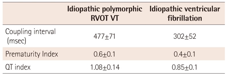
The term “idiopathic polymorphic RVOT VT” should be reserved for patients with frequent and otherwise typical outflow tract ventricular ectopy (see “clinical characteristics” below) who develop rapid polymorphic VT as their only sustained arrhythmia or (not infrequently) in addition to well-tolerated sustained monomorphic VT (Fig. 1, 2).8) This definition recognizes the fact that ECG provides only an approximate estimate of the site of origin of an arrhythmia, and that detailed intracardiac mapping might ultimately localize the site of origin of the arrhythmia to the LVOT or contiguous structures. The term “idiopathic” indicates that organic heart disease has been excluded. However, as discussed in detail elsewhere,12) differentiating a truly idiopathic RVOT VT from RVOT VT related to less-than-overt forms of right ventricular dysplasia is not always straightforward. Finally, this definition assumes that idiopathic polymorphic RVOT VT and idiopathic VF13) are different diseases. Importantly, in 14% of cases of idiopathic VF, the arrhythmia originates in the RVOT,14) and several ECG characteristics differentiate patients with RVOT-vrelated idiopathic VF from the more common form of idiopathic VF originating from Purkinje fibers.14) Specifically, the coupling interval of extrasystoles triggering idiopathic VF from the RVOT, although “short,” is not as short as the ultra-short coupling interval of typical (Purkinje-related) idiopathic VF9) (355±30 vs. 280±26 msec, p=0.01).14) In addition, RVOT extrasystoles are wider than the Purkinje-related ectopic beats (145±12 vs. 126±18 msec, p=0.04).14) As opposed to patients with idiopathic VF originating from the RVOT, who usually have cardiac arrest as their presenting symptom (Fig. 4) and who have little-to-no ventricular ectopic activity between arrhythmic events, patients with idiopathic RVOT VT have a long history of palpitations and have frequent RVOT extrasystoles (often with documented episodes of well-tolerated monomorphic VT) before they actually develop polymorphic VT (Fig. 2).8)10)15)
Incidence
In a retrospective series involving 91 patients referred over the course of five years to a single Japanese center for ablation of VT originating from the RVOT in the absence of heart disease, 14 patients (15%) had documented polymorphic VT, including four (4%) with VF.16) Because of recall bias and referral bias, this series probably overestimated the risk of polymorphic VT among patients with RVOT VT. Furthermore, the incidence of polymorphic VT among patients presenting with ventricular extrasystoles (rather than with RVOT VT, as in this series) would be drastically smaller. A different series, involving 130 patients evaluated for ventricular arrhythmia in the absence of organic heart disease, presented a more realistic number, wherein four patients (3%) had documented polymorphic VT originating from the RVOT or cardiac arrest with VF.17) Since our original description of this phenomenon one decade ago, we have not encountered additional patients presenting with idiopathic RVOT extrasystole or RVOT monomorphic VT who have gone on to develop polymorphic VT. Clearly, malignant polymorphic RVOT VT is rare, and the great challenge is to distinguish the small minority of patients with this malignant disease from the majority of patients with benign idiopathic RVOT arrhythmia.
Clinical Characteristics
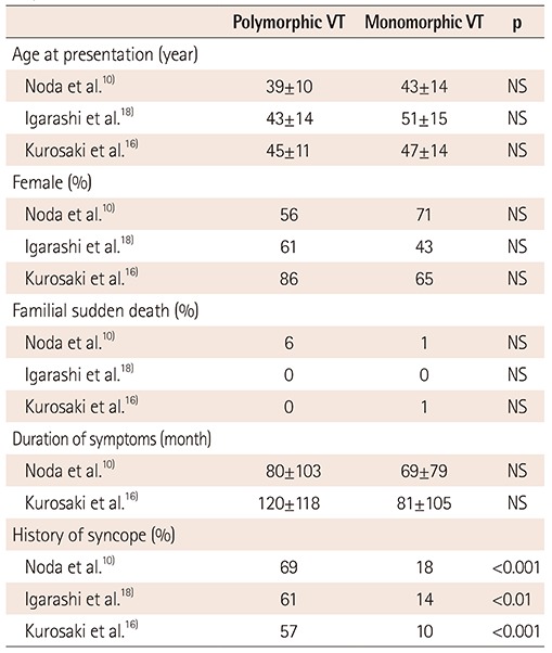
A typical patient with malignant RVOT VT is an otherwise healthy adult presenting with a long history of palpitations. In different case series, the mean age of patients at the time of presentation with polymorphic VT is 39-45 years.10)16)18) Females predominate, representing 56% to 85% of patients in different series.10)16)18) A family history of sudden death is reported only exceptionally.10)18) In general, patients with polymorphic VT report a history of palpitations beginning 6-10 years prior to the malignant arrhythmic event. In all of these aspects, patients with malignant polymorphic RVOT VT are not different from patients with the more common and benign form of monomorphic RVOT VT. Not surprisingly, a history of syncope is reported more frequently among patients with polymorphic VT (Table 1).
Electrocardiographic Characteristics
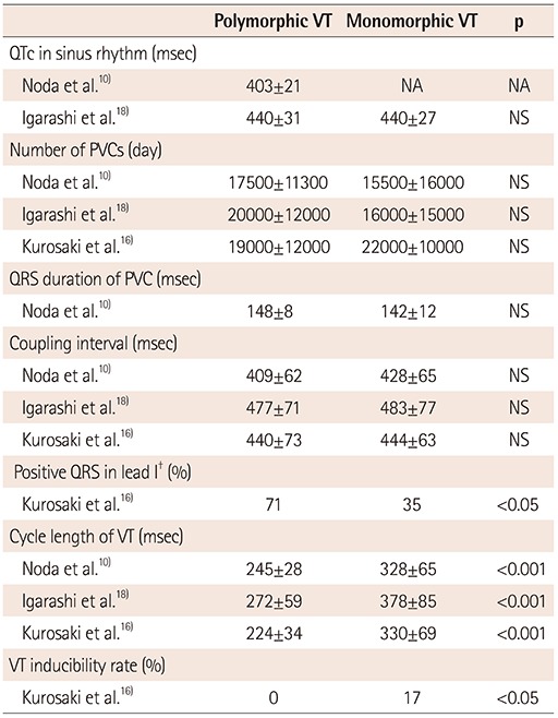
The baseline ECG during sinus rhythm, both for patients with the common benign form of RVOT monomorphic VT and for patients with the rare form of polymorphic RVOT VT, is strictly normal. To date, neither “early repolarization”19)20) nor QT intervals in the low range of normal21) (two ECG characteristics associated with idiopathic VF)19)20)21) have been reported in patients with polymorphic RVOT VT. In fact, the mean QTc of patients with polymorphic VT is 400-440 msec (values representing the 30th-90th percentiles of QTc in the healthy population),22) which is not different from the QTc of patients with monomorphic RVOT VT (Table 2).18)
Coupling interval
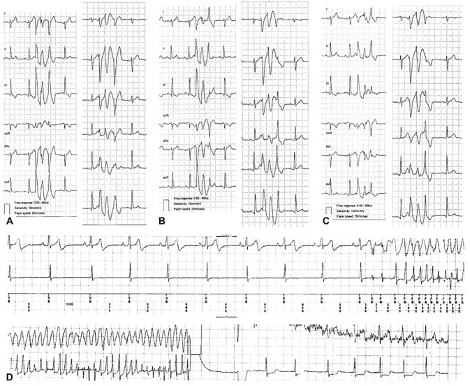
As mentioned, the coupling interval of the extrasystole-initiating polymorphic VT events is short in some patients, but is not short in all patients. Hence, the mean coupling interval of extrasystoles initiating polymorphic VT is not significantly shorter than the mean coupling interval of extrasystoles initiating monomorphic VT and is not different from the coupling intervals of extrasystoles that do not trigger arrhythmias (Table 2) (Fig. 5B). Also, RVOT extrasystoles with varying coupling intervals have been associated with an increased risk of polymorphic VT.23)
Prematurity index
In one series comparing 42 episodes of polymorphic VT and 48 episodes of monomorphic VT from the RVOT, the basic sinus rate of patients was slower immediately prior to the onset of polymorphic VT (Fig. 5).18) Consequently, despite similar coupling intervals, the prematurity index (defined as the ratio of the coupling interval of the first VT beat or isolated extrasystoles to the preceding R-R interval during sinus rhythm) was shorter prior to the onset of polymorphic VT. Similarly, the QT index (defined as the ratio of the coupling of the first VT beat or isolated premature vetricular contraction to the QT interval of the preceding sinus complex) was shorter during polymorphic VT (Fig. 5).18) Among these patients, a prematurity index <0.73 predicted a polymorphic arrhythmia in the event of VT with good sensitivity but poor specificity (91% and 44%, respectively).18) Of note, these values are longer than the values we have previously reported for patients with idiopathic VF (Table 3).9)
Site of origin
Kurosaki et al.16) were the first to note that polymorphic VT episodes are frequently initiated by RVOT extrasystoles with positive QRS morphology in lead I (Table 2). This intriguing ECG characteristic is also observed in case reports.24)25)26)27)28) Intracardiac mapping of the RVOT has located these extrasystoles with positive QRS in lead I to the posterior aspect of the RVOT.28)
Mechanism
In general, idiopathic RVOT arrhythmias are believed to be due to triggered activity.29)30) As discussed in detail elsewhere,31) initiation of polymorphic VT/VF, which is triggered by an ectopic beat with a short coupling interval, could well represent the “normal” response to a timed electrical stimuli falling on the “vulnerable phase” of the ventricle at a point when the dispersion of ventricular refractoriness is greatest.32) Explaining the initiation of polymorphic VT as an extrasystole falling after the end of the T-wave of the preceding sinus beat is more difficult, and we can only speculate about potential mechanisms. One possibility is that the polymorphic VT/VF is triggered by a rapid series of delayed after-depolarizations (analogous to the experimental induction of VF by a series of rapid electrical stimuli), in which the first after-depolarization is concealed, but lowers the fibrillation threshold for subsequent stimulus.33) A second possible mechanism is that the polymorphic arrhythmia is induced not by the first, but instead by the second ventricular extrastimulus, which invariably has a very short coupling interval.11)16) Indeed, there are examples of polymorphic VT beginning after a long-coupled RVOT extrasystole that is followed by a short-coupled beat originating from Purkinje fibers at the moderator band. In such an example, the second (moderator band) extrasystole is the real trigger of the polymorphic VT (F. E. Marchlinski, personal communication). A third possibility is that the RVOT ectopic activity leading to VF could be a manifestation of modulated parasystole and reflection (a term used to denote a parasystolic pacemaker undergoing slow diastolic depolarization, which has been triggered to fire prematurely by the electrical activity of adjacent tissue).34) The observation that varying coupling intervals correlate with a higher risk of polymorphic VT23) is consistent with this last possibility.
Identification of Patients at Risk
Recognizing the small proportion of patients at risk for potentially lethal polymorphic RVOT VT among the large majority of patients with benign RVOT extrasystoles is a formidable challenge.11) The only clinical characteristic that specifically increases the odds of having polymorphic VT is a history of syncope (Table 1). Certainly, patients with RVOT extrasystoles should be suspected of having polymorphic VT when their clinical history is consistent with a malignant syncope. On the other hand, both benign vagal syncope and benign idiopathic RVOT arrhythmias are common, and it is not unusual to encounter patients who have both—unrelated—medical conditions. It could be argued that patients with RVOT extrasystoles who have syncope that appears to be benign vagal syncope on clinical grounds should be regarded as patients with asymptomatic (i.e., unrelated) extrasystoles. Here is where the challenge becomes extreme,11) because, as reported by Kataoka et al.,28) an otherwise benign episode of spontaneous vagal syncope can unpredictably trigger polymorphic RVOT VT in a prone patient (Fig. 3B). For the asymptomatic patient, documentation of short-coupled extrasystoles probably calls for an aggressive approach with radiofrequency ablation (duly admitting that there is no data about the natural history of short-coupled extrasystoles).
Natural History and Therapy
Patients with documented RVOT polymorphic VT are treated with radiofrequency ablation of any and all ectopic beats that resemble arrhythmia triggers.10)16)18) Whether or not all of these patients should also undergo implantation with an implantable cardioverter-defibrillator (ICD) is an open question, because little is known about the rate of recurrence after successful ablation. Of note, the five-year recurrence of malignant arrhythmias following successful ablation of idiopathic VF is as high as 38%.35) Comparable long-term figures are not available for RVOT polymorphic VT. Similarly, quinidine is extremely effective for preventing recurrences in idiopathic VF,1)13) but there is no data on the efficacy of quinidine in polymorphic RVOT VT. Non-inducibility of VT at the end of an ablation procedure cannot be used as evidence of success, because patients do not necessarily have inducible VT at baseline (Table 2). Finally, little is known about the natural history of patients with documented idiopathic RVOT polymorphic VT. Interestingly, two of our initial patients remained untreated (except for ICD implantation in both patients).8) One of these patients has remained free of arrhythmias following a cardiac arrest 12 years prior, whereas the other patients has remained asymptomatic, but has documentation of non-sustained polymorphic VT 18 years after her initial arrhythmic event (Fig. 1D). Looking at these patients, we can only wonder if the RVOT polymorphic VT is simply a matter of “bad luck.” That is, can the phenomenon be characterized as an otherwise innocent extrasystole with extremely bad timing (occurring in the vulnerable phase of the cardiac cycle).
 XML Download