

 PDF
PDF ePub
ePub Citation
 Print
Print


Introduction
Cardiac ischemic post-conditioning (PostC) is the technique wherein alternating cycles of sub-lethal myocardial ischemia and reperfusion are applied after a sustained insult. It is a cardioprotective strategy that can reduce reperfusion injury. Infarct size reduction and improvements in left ventricular ejection fraction have been demonstrated with mechanical or pharmacological postC after spontaneous acute myocardial infarction.1) Various pathways have been reported during ischemic postC such as reperfusion injury salvage kinase pathways (p44/42-ERK1/2/PI3k/AKT) as well as survival activating factor enhancement pathways (TNF-α, JAK/STAT).2)3) Hypercholesterolemia (HC) has been reported to abrogate the beneficial effects of postC in ischemia-reperfusion (I/R)-induced myocardial injury 4. It has been reported that HC abolishes the benefits of postC by alterations in cytochrome c, Akt, ERK1/2, caspase 9, caspase 3, nitric oxide synthase, endothelial nitric oxide synthase (eNOS), mitochondrial adenosine triphosphate (ATP)-dependent K+ channel, Bcl-2, Bax and PI3K/Akt/eNOS pathways.4)5)
HC activates the renin-angiotensin system, increases angiotensin II type 1 (AT1) receptor density and functional responsiveness of AT1 receptors.6) The angiotensin II type 1 receptor antagonist protects the heart against acute I/R injury.7) Furthermore, AT1 blockers have been reported to provide anti-inflammatory, anti-oxidative, cardio-protective, anti-hypertensive and beneficial effects in I/R myocardial injury.7)8)
Azilsartan is a new AT1 blocker that has been recently launched; nothing much is known about its utility in I/R myocardial injury, ischemic PostC of the heart and HC. The present study has been designed to investigate for the first time, the utility of a novel AT1 blocker, azilsartan in I/R injury and ischemic PostC of the heart of normal and hypercholesterolemic rats.
Materials and Methods
Animals
Male Wistar rats (weight 250 to 300 g) were obtained from Henan University Animal Public Service Centre (Henan, China). All animals were treated in accordance with the Guide for the Care and Use of Laboratory Animals and approved by the Research Review and Ethics board of Henan University Huaihe Hospital, Henan, China (Ref no. 20131110, dated 10 Nov, 2013). All the animals were housed at a temperature of 23℃, 60% humidity, and 12 hlight/dark cycles. Water and food were freely available to the animals.
Drugs and chemicals
All the chemicals and reagents were of AR grade and prepared freshly before use.
Induction of HC in rats
Male Wistar rats (250-300 g) were employed in the present study. Experimental HC was induced in the animals by changing their diet with that of a high-fat diet for 8 weeks.9) The establishment of hyperlipidemia was confirmed by serum lipid profiling of the animals.
Isolated rat heart preparation
The rats were anesthetized with pentobarbital sodium (100 mg/kg intraperitoneally). The heart was immediately isolated and mounted on a Langendorff apparatus.4)5) A circulating water jacket was provided to surround the heart for temperature maintenance at 37℃. The preparation was perfused with Krebs Henseleit solution, pH 7.4, maintained at 37℃, and bubbled with 95% O2 and 5% CO2. The perfusion pressure was maintained at 80 mm of Hg for the maintenance of a coronary flow rate at around 7 mL/min. For induction of global ischemia, the inflow of Krebs Henseleit solution was blocked for 30 min, and then reperfusion for 120 min was re-established by allowing the inflow of Krebs Henseleit solution.10) Left ventricular hemodynamic parameters were measured via a latex balloon inserted in the left ventricle. The balloon catheter was linked to a pressure transducer connected to the physiological signal acquisition system (PowerLab, AD Instruments, Shanghai, China) to monitor the contractile function. To induce the PostC we have utilized six cycles of 10 sec ischemias and 10 sec reperfusions, after the 30 min ischemia and before the 120 min reperfusion. This is an acceptable and widely used protocol for induction of PostC during ischemia and reperfusion injury of the isolated heart.4)5)
Experimental protocol
In the present study, a total of eleven groups were employed (n=12) as per the experimental protocol (Fig. 1). All the isolated Langendorff-perfused rat hearts were first allowed to stabilize with the help of a perfused Krebs Henseleit solution for 10 min.
Group I-sham control–normocholesterolemic (NC)
The isolated rat heart was perfused with the Krebs Henseleit solution for 200 min.
Group II-I/R-NC
The isolated rat heart was exposed to global ischemia for 30 min, followed by reperfusion for 120 min with a Krebs Henseleit solution.
Group III-ischemic-post-conditioned-NC
The isolated rat heart was exposed to 30 min of global ischemia, followed by six cycles of 10 sec global ischemias and 10 sec reperfusions, to establish ischemic-PostC in the heart. After that, the heart was reperfused for 120 min with a Krebs Henseleit solution.
Group VI-sham control-HC
The isolated rat heart was exposed to 30 min of global ischemia followed by six cycles of 10 sec global ischemias and 10 sec reperfusions, to establish PostC in the heart. After that the heart was reperfused for 120 min with an azilsartan (5 mM) solution.
Group VI-sham control-HC
The heart was isolated from hypercholesterolemic and the remainder of the procedure was the same as that of group I.
Group VII-I/R-HC
The heart was isolated from hypercholesterolemic rat and the remainder of the procedure was the same as that of group II.
Group VIII-ischemic post-conditioned-HC
The heart was isolated from hypercholesterolemic rat and the remainder of the procedure was the same as that of group III.
Group IX-I/R-azilsartan post treatment-HC
The heart was isolated from hypercholesterolemic rat and the remainder of the procedure was the same as that of group IV.
Group X–ischemic post-conditioned-azilsartan post treatment-HC
The heart was isolated from hypercholesterolemic rat and the remainder of the procedure was the same as that of group V.
Group XI-ischemic post-conditioned-L-N5-(1-Iminoethyl)ornithine (L-NIO) hydrochloric acid (HCI) azilsartan post treatment-HC
The heart was isolated from hypercholesterolemic rat and perfused with a Krebs Henseleit solution for 10 min. After that, the heart was exposed to 30 min of global ischemia followed by six cycles of 10 sec global ischemias and 10 sec reperfusions, to PostC in the heart. After that the heart was reperfused for 15 min with L-NIO hydrochloride (1 mM) followed by reperfusion for 105 min with an azilsartan (5 mM) solution.
Estimation of lactate dehydrogenase, creatine kinase, tumor necrosis factor-alpha, troponin I and nitrite in coronary effluent
To assess the myocardial injury, the release of lactate dehydrogenase (LDH) (0 and 30 min) and creatine kinase (CKMB) (5 min) in the coronary effluent was estimated with the help of commercially available enzymatic kits (Bangjing, Shanghai, China). The concentrations of tumor necrosis factor-alpha (TNF-α) (1, 5, and 10 min), TnI (60 min) were measured by the sandwich ELISA technique (R&D system, Minneapolis, MN, USA) and quantified photometrically, at an absorbance of 450 nm. Furthermore, nitric oxide levels were measured (spectrophotometrically at 550 nm) as the nitrite concentration (0 and 15 min) uses Greiss reagent.13)
Assessment of oxidative stress
The left ventricle was homogenized in an ice-cold phosphate buffer 0.05 M (pH 7.4) and the clear supernatant was utilized for the spectrophotometric measurement of thiobarbituric acid reactive substances (TBARS),16) superoxide anion (SA)17) and reduced form of glutathione (GSH)18) at 532 nm, 540 nm and 412 nm respectively, as per the previously published reports.
Results
Effect of high-fat diet on serum lipi
The administration of a high fat diet has significantly increased the total cholesterol (260 mg/dL), triglycerides (210 mg/dL) and low-density lipoproteins (15 mg/dL) along with a significant reduction of high-density lipoproteins (30 mg/dL) as compared to the control animals (76 mg/dL; 63 mg/dL; 32 mg/dL; 45 mg/dL) on a normal chow diet.
Effect on hemodynamic parameters of heart
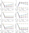
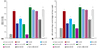
When compared to sham control, 30 min global ischemia and 120 min reperfusion in NC and HC animals have significantly enhanced left ventricular end-diastolic pressure along with a significant reduction in heart rate, left ventricular developed pressure, ±dP/dt max and coronary flow rate. I/R-injury induced impairment of hemodynamic parameters of hearts was attenuated by ischemic-PostC (six cycles of 10 sec ischemias and 10 sec reperfusions) and azilsartan post treatment in NC rats. HC has significantly abolished the beneficial effects of PostC on hemodynamic parameters. The perfusion of azilsartan to the heart of HC-ischemic post-conditioned animals has significantly normalized the beneficial effect of PostC on the hemodynamic parameters of heart, which were significantly inhibited by the L-NIO, a potent inhibitor of eNOS (Fig. 2).
Effect on the release of lactate dehydrogenase, creatine kinase and tumor necrosis factor-alpha
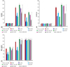
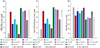
When compared to sham control, 30 min global ischemia and 120 min reperfusion in NC and HC animals have significantly enhanced LDH (at 0 min and 30 min of reperfusion), CK-MB (at 5 min of reperfusion) and TNF-α (at 1 min and 5 min of reperfusion). I/R-injury induced impairment in LDH, CK-MB, and TNF-α levels was attenuated by ischemic-PostC (six cycles of 10 sec ischemia and 10 sec reperfusion) and azilsartan post treatment in NC rats. HC has significantly abolished the beneficial effects of PostC on LDH, CK-MB and TNF-α levels. The perfusion of azilsartan to the heart of HC-ischemic post-conditioned animals has significantly normalized the beneficial effect of PostC on the LDH, CK-MB and TNF-α levels, which were significantly inhibited by the L-NIO, a potent inhibitor of eNOS (Fig. 3).
Effect on the release of nitrite and troponin I
When compared to sham control, a 30 min global ischemia and 120 min reperfusion in NC and HC animals have significantly reduced nitrite (at 0 min and 15 min of reperfusion), and troponin I (TnI) levels (at 60 min of reperfusion). I/R-injury induced impairment in nitrite and TnI levels, was attenuated by ischemic-PostC (six cycles of 10 sec ischemias and 10 sec reperfusions) and azilsartan post treatment in NC rats. HC has significantly abolished the beneficial effects of PostC on nitrite and TnI levels. The perfusion of azilsartan to the heart of HC-ischemic post-conditioned animals has significantly normalized the beneficial effect of PostC on the nitrite and TnI levels, which were significantly inhibited by the L-NIO, a potent inhibitor of eNOS (Fig. 4).
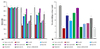
Effect on the % infarct size and left ventricle collagen content
When compared to sham control, a 30 min global ischemia and 120 min reperfusion in NC and HC animals have significantly enhanced the percentage of the infarction size and left ventricle collagen content of rat hearts. I/R-injury induced impairment in the percentage of the infarction size and left ventricle collagen content was attenuated by ischemic-PostC (six cycles of 10 sec ischemias and 10 sec reperfusions) and azilsartan post treatment in NC rats. HC has significantly abolished the beneficial effects of PostC on the percentage of the infarction size and left ventricle collagen content. The perfusion of azilsartan to the heart of HC-ischemic post-conditioned animals has significantly normalized the beneficial effect of PostC on the percentage of the infarction size and left ventricle collagen content, which were significantly inhibited by the L-NIO, a potent inhibitor of eNOS (Fig. 5).
Effect on the heart oxidative stress
When compared to sham control, a 30 min global ischemia and 120 min reperfusion in NC and HC animals have significantly enhanced the heart's oxidative stress (noted as a significant increase in TBARS and SA along with significant reduction in GSH levels of the heart). I/R-injury enhanced oxidative stress, was attenuated by ischemic-post-conditioning (six cycles of 10 sec ischemias and 10 sec reperfusions) and azilsartan post treatment in NC rats. HC has significantly abolished the beneficial effects of PostC on oxidative stress. The perfusion of azilsartan to the heart of HC-ischemic PostC animals has significantly normalized the beneficial effect of PostC on oxidative stress, which was significantly inhibited by the L-NIO, a potent inhibitor of eNOS (Fig. 6).
Discussion
I/R induced injury of heart
In the present study, a 30 min ischemia and 120 min reperfusion significantly induced myocardial injury, when compared to sham control animals. I/R myocardial injury has been shown to have a direct correlation of infarct size with that of cardiac hemodynamic parameters, release of LDH, CK-MB, TnI and inflammatory markers like TNF-α, in the coronary effluent after an ischemic insult to the myocardium.9)14)19) LDH release was found to be maximum at 0 min, i.e. immediately after the reperfusion. On the other hand CK-MB and TNF-α release was found to be maximum at 5 min intervals.
High level of oxidative stress in I/R is evident by significantly higher levels of TBARS, and SA, along with significantly lower levels of GSH, was found in the heart exposed to I/R injury. TNF-α is a specific marker of inflammation and its higher levels in the coronary effluent signifies the high level of inflammation during I/R injury. Lower levels of nitrite are an index of decreased availability of NO which may result in cardiac damage.13)
Higher levels of collagen content in the left ventricle signify a possible cardiac death during I/R injury of the heart. All the above alterations have resulted in increased myocardial injury and death, which has resulted in increased infarct size and consequent reduction in the coronary flow rate during an I/R-induced myocardial injury. These findings are in accordance with the previously published reports.9)13)14)
Beneficial effect of ischemic PostC in I/R-injury of heart
Six cycles of 10 sec ischemias and 10 sec reperfusions have post-conditioned the normal rat heart against the myocardial injury during the I/R insult of a 30 min ischemia and 120 min reperfusion. PostC has markedly attenuated the I/R myocardial injury in normal rat hearts and the results are in parallel to previous studies.4)5) Cardiac ischemic post conditioning-the technique of applying alternating cycles of sub-lethal myocardial ischemia and reperfusion after a sustained insult-is one cardioprotective strategy that can reduce reperfusion injury. Infarct size reduction and improvements in left ventricular ejection fraction have been demonstrated with mechanical or pharmacological PostC, both after a spontaneous acute myocardial infarction, and associated with cardiac surgery.1) Badalzadeh et al.10) have reported that I/R injury may mitochondrial ATP-dependent potassium (mito-KATP) channels and nitric oxide system as the main players in the induction of a myocardial injury. In our study, the disturbance in the nitric oxide system is evident as there was a reduction of nitrite release in the coronary effluent after an ischemic insult. It has been suggested that during an I/R injury of the heart, activation of IkB and NFkB occur.20) These are the important modulators of inflammation; high levels of inflammations are evident from the increase in the release of TNF-α in this study.
HC abolished benefits of ischemic-PostC in I/R injury of heart
Administration of a high-fat diet rich in cholesterol to the rats for 8 weeks had markedly enhanced the total serum cholesterol, triglycerides and low-density lipoproteins along with the reduction of beneficial lipids, and high-density lipoproteins. Thus, a high-fat diet has successfully established HC as well as hyperlipidemia in the rats. When the hearts of hypercholesterolemic rats were exposed to the PostC, PostC achieved very limited protection in I/R myocardial injury. HC has abolished PostC induced correction of hemodynamic parameters, reduction in the infarct size, release of LDH, CK-MB, TNF-α, TnI, left ventricle collagen content, TBARS, SA along with the increase of coronary flow rate, nitrite release, and GSH levels. This suggests that HC has significantly abolished all the benefits of PostC in I/R injury of hypercholesterolemic rats heart, which is in parallel to previous studies.4)5) It has been suggested that HC may induce excessive apoptosis by down-regulating Bcl-2 and upregulating Bax, cytochrome c, caspase 9 and caspase 3 along with inhibition of phosphorylation of Akt and ERK1/2. These may ultimately result the inactivation of RISK signal pathways and dysregulation of downstream apoptosis-related pathway.5) Involvement of Rho kinase, PI3K/Akt/eNOS signal pathways have also been reported in HC-induced impairment of PostC benefits during I/R-injury of heart.4) Very little work has been done on this aspect and thus much research work is required to unearth the other possible mechanisms of PostC during HC.
Benefits of AT1 blocker in I/R injury and ischemic-post-conditioning in normal as well as hypercholesterolemic heart: role of eNOS
The perfusion of the novel AT1 receptor inhibitor, azilsartan post treatment has significantly protected the I/R induced injury of heart in normocholesterolemic animals, in a similar manner as that of PostC mediated protection in I/R injury of normal animals. Furthermore, perfusion of azilsartan in hypercholesterolemic-ischemic post-conditioned animals has significantly reversed the HC induced inhibition of the beneficial effects of PostC. In I/R myocardial injury and hypercholesterolemic-ischemic post-conditioned animals, perfusion of azilsartan has significantly corrected the hemodynamic parameters of the heart, reduced the release of LDH, CK-MB, TNF-α, TnI nitrite in coronary effluent, oxidative stress, left ventricle collagen content, infarct size along with significant correction of coronary flow rate. Previously various in-vitro doses of azilsartan have been utilized such as 1 mM 11 and 0.005% (-3 mM).12) In our preliminary studies, we have first utilized1)3) and 5 mM/L doses of azilsartan. The significant protection was observed with 5 mM/L doses (data not shown), which was then selected for the protocol of the present study.
To understand the exact mechanism behind the beneficial effects of azilsartan (a novel AT1 receptor antagonist), on PostC in hypercholesterolemic rat heart, we have employed the L-NIO, which is a potent inhibitor of eNOS. The beneficial effects of azilsartan perfusion on PostC in hypercholesterolemic rat heart was significantly inhibited by the perfusion of L-NIO which suggests that azilsartan has provided beneficial effects on PostC in hypercholesterolemic rat heart by specific modulation of eNOS.
Azilsartan is an angiotensin receptor blocker which is recently approved for treating patients with hypertension, introduced in the year 2011, which exerts its benefits predominantly through AT1 receptors.21) It has been reported that azilsartan provided kidney and heart protective effects due to its ability to lower blood pressure and improve endothelial functions.22) Iwanami and colleagues23) have reported that the hypotensive and anti-hypertrophic effects of azilsartan may involve activation of the ACE2/Ang-(1-7)/Mas axis with AT1 receptor blockade. The azilsartan has been shown to exert favorable biological effects on the hearts during left ventricular pressure overload in obese insulin-resistant conditions.24) In vitro studies showed that azilsartan medoxomil was hydrolyzed rapidly to azilsartan in plasma, hepatic S9 fractions, and intestinal S9 fractions from all species tested. In human hepatic microsomes, azilsartan was further decarboxylated to the pharmacologically inactive metabolite M-I by the cytochrome P450 enzyme (CYP) 2C8, or was O-dealkylated to the inactive metabolite M-II by CYP2C9. The half maximal inhibitory concentration (IC50) values for azilsartan medoxomil for the in vitro inhibition of human hepatic CYP isoforms, CYP2C8, CYP2C9, CYP3A4, CYP2B6, CYP1A2, and CYP2C19 ranged from 3.5 to 66 µmol/L.25)
Not much is known about the utility of azilsartan in I/R-induced myocardial injuries, PostC, and HC. Sgarra et al.26) have recently reported that AT1 blocker may provide benefits similar to PostC of theheart. Azilsartan is an angiotensin receptor blocker that has been recently approved for treating patients with hypertension. Introduced in the year 2011, it exerts its benefits predominantly through blockade of AT1 receptors.21) Not much is known about the utility of azilsartan in I/R-induced myocardial injuries, PostC, and HC.
Azilsartan has been reported to provide protection against oxidative stress and inflammation. Furthermore, it has also been found that azilsartan improves lipid profiles, heart function, and endothelial function as well.22) It has been suggested that azilsartan decreases TNF-α expression better than candesartan cilexetil.27) Non-hypertensive doses of azilsartan have been reported to cardiac remodeling, infarct size and fibrotic changes after induction of myocardial infarction.28) It has been reported earlier that, eNOS dysfunction is one of the important pathways during myocardial I/R-injury.29) Wu et al.4) have reported that PostC provides benefits in I/R-injury of heart by upregulation of eNOS. HC has been reported to abolish the beneficial effects of PostC during I/R-injury of heart by impairing eNOS function.4) AT1 blockers have been reported to enhance eNOS expression.30) Matsumoto et al.12) have reported that azilsartan treatment corrects the phosphorylation of eNOS. Thus, azilsartan, a novel AT1 blocker may have provided its benefits due to its anti-oxidative stress, cardioprotective, anti-inflammatory, and nitric oxide-releasing effects via modulation of eNOS.
Conclusions
The results of this study suggests following aspects of azilsartan, which are unique to this study: 1) azilsartan significantly provides protection in I/R-induced myocardial injuries in normocholesterolemic rats, similar to the benefits provided by PostC in I/R-induced myocardial injuries in normocholesterolemic rats; 2) azilsartan has also re-established the beneficial effects of PostC in I/R myocardial injuries, abolished by HC; 3) azilsartan has also provided its beneficial effects through specific modulation of eNOS. Further, research on azilsartan is warranted to unearth the various other mechanisms involved in the beneficial effects in I/R injury and PostC of the heart.
 XML Download