

 PDF
PDF ePub
ePub Citation
 Print
Print


Introduction
The left atrial appendage is regarded as just a minor structure added to the left atrium. However, it has been recently reported that the left atrial appendage plays a hemodynamic role for atrial function as well being the major source of the thrombus.1)2) During the embryonic period, the left atrial appendage is developed from the left wall of the primary atrium and has anatomical as well as a physiological distinction from the left atrium proper.
Recently interest in the left atrial appendage has increased due to the importance of its evaluation before a catheter ablation or electrical cardioversion for atrial fibrillation. The transesophageal echocardiography and computed tomography have provided clear imaging of the left atrial appendage.
Case
Case 1
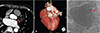
A 68-year-old man having no history of cardiac surgery was referred for a catheter ablation for drug-refractory atrial fibrillation. He had taken 8 mg of candesartan and 25 mg of hydrochlorothiazide for hypertension. He also had diabetes without the usage of an oral hyperglycemic agent. He suffered from palpitation and chest discomfort for 6 months, and started anticoagulation for catheter ablation one month prior. Transthoracic echocardiography revealed moderate mitral regurgitation and significant left atrial enlargement (AP diameter=48 mm, volume=116 mL). He underwent cardiac computed tomography (CT), in which the left atrial appendage was not observed (Fig. 1A, B). In a 3D reconstructed image, the left atrial appendage was not observed. However, the right atrial appendage was observed and its morphology and site were normal; the four pulmonary veins also looked normal. Pro B-natriuretic peptide was within normal limits (145.8 pg/mL) and A left atrial angiogram also revealed the absence of the left atrial appendage (Fig. 1C). A catheter ablation was performed and the initial rhythm was sinus. However, rapid atrial pacing induced cavotricuspid isthmus dependent atrial flutter requiring intracardiac electrical cardioversion (3J). Then, pulmonary vein isolation with antral level and cavotricuspid ablation were successfully achieved. Isoproterenol infusion (up to 10 mcg/min) also did not provoke atrial fibrillation or flutter. For 16 months, the patient had not complained of any symptoms and sinus rhythm was maintained with an antiarrhythmic drug (flecainide 50 mg bid). Warfarin was discontinued 12 months after the catheter ablation. Follow up echocardiography showed a decreased left atrialdecreased left atrial diameter (48 mm to 4 mm).
Case 2
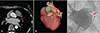
A 58-year-old woman presented with drug-refractory atrial fibrillation. Three months ago, she had undergone catheter ablation for typical atrial flutter. She was taking anti-hypertensive medication and a brain magnetic resonance imaging revealed an old lacunar infarct. Echocardiography showed normal left ventricular function and left atrial enlargement (AP diameter: 45 mm). She underwent cardiac CT, in which only the stump of the left atrial appendage was observed (Fig. 2A, B). Left atrial angiogram was performed but the small ectopic appendage could not be found (Fig. 2C). Electrophysiologic study confirmed a bidirectional block at the carvotricuspid isthmus. Pulmonary vein isolation was done and an atrial arrhythmia was unable to be induced by rapid atrial pacing (cycle length 170 ms). Normal sinus rhythm was maintained without the use of an anti-arrhythmic drug and there were no incidences of stroke. Rivaroxaban was used for anticoagulation.
Discussion
The left atrial appendage usually develops within four weeks of the embryonic period. It secretes atrial natriuretic factor,7) acts as a left atrial reservoir, and has contractile functions.8) Above all things, intracardiac thrombus in atrial fibrillation was mostly observed in the left atrial appendage. The physiological and clinical consequences of aplastic left atrial appendage are unknown.
In case of the absence of the left atrial appendage during transesophageal echocardiography, flush thrombus and a poor echocardiographic window could be considered as differential diagnoses. CT is very helpful to confirm the absence of the left atrial appendage as in the present case.
Brain natriuretic peptide is produced and stored in both the atria and appendage, with secretion proportional to hemodynamic balance.9) More densely granule cells occur more often in the right than left atrial appendage.7) A study demonstrated that bilateral atrial appendectomy decreased plasma atrial natriuretic peptide levels.10) In the present case, the patient seemed to have normal secretion of natriuretic peptides and fluid status despite the absence of a left atrial appendage.
Regarding the ablation technique, cautious catheter manipulation around left atrial ridge should be necessary in case of an aplastic left atrial appendage. It is possible that atrial tissue around the left atrial ridge is different with usual one, for example so friable or thick. Fortunately, left pulmonary vein isolation was easily achieved.
There is no guideline for anticoagulation in case of the aplastic left atrial appendage. Although our patient had two risk factors for embolic risk (diabetes and hypertension) and was relatively old age (68 years old), warfarin was changed to aspirin one year after the catheter ablation because there was no recurrence of atrial fibrillation for one year and reverse atrial remodeling was observed.
In conclusion, aplastic left atrial appendage is not a common congenital anomaly and cautious catheter manipulation is necessary. Further study is required for the needs of anticoagulation in aplastic left atrial appendage patients in the future.
 XML Download