

 PDF
PDF ePub
ePub Citation
 Print
Print


Introduction
Kawasaki disease (KD), first described in 1967,1) is an acute self-limited systemic vasculitis of unknown etiology. It occurs predominantly in children under 5 years of age and results in coronary artery abnormalities in 15-25% of untreated children.1)2)3)4) Specific diagnostic tools for KD are unavailable, hence, the diagnosis is usually based on principal clinical features of fever, bilateral non-exudative bulbar conjunctivitis, erythema of the lips and oral mucosa, changes in the extremities, skin rash, and cervical lymphadenopathy. Among the principal diagnostic criteria for KD, cervical lymphadenopathy is the least common, occurring in approximately 24-75% of patients with KD.5)6) A few KD patients present with only fever and cervical lymphadenopathy before other clinical features appear.3)5)7)8) KD patients with cervical lymphadenopathy as the dominant manifestation have significant risks for misdiagnosis as bacterial cervical lymphadenitis or other lymphadenopathies, delay in diagnosis and appropriate treatment, and development of coronary complications.7)9)10)11) Consequently, characterization of patients with KD presenting with only fever and cervical lymphadenopathy before other clinical signs appear may help reduce the development of cardiac complications. However, whether KD patients with only fever and cervical lymphadenopathy at first presentation have an independent risk for intravenous immunoglobulin (IVIG) resistance or coronary artery lesions (CALs) is unclear.6)9) The purpose of the present study was to characterize the clinical features of these unusual KD patients, as compared with those of other KD patients, to determine whether this is a severe form of KD with increased risks of IVIG resistance and CALs.
Subjects and Methods
Patients
The medical records, echocardiography data, and laboratory parameters of 146 patients diagnosed with KD and treated at Konkuk University Hospital between May 2009 and December 2013 were reviewed retrospectively. KD was diagnosed according to the criteria of the American Heart Association, 2004.2)
The diagnostic criteria for KD were as follows: fever with a duration ≥3 days but ≤10 days, at least 4 of 5 principal criteria for KD i.e., conjunctival injection, skin rash, oral mucosal changes, peripheral extremity changes, and cervical lymphadenopathy; 2 or 3 principal criteria plus coronary artery abnormalities documented by echocardiography; or 2 to 3 criteria with elevated inflammatory indices and 3 supplemental laboratory criteria.
Patients with KD were classified into two groups according to initial clinical manifestations. KD patients with only fever and cervical lymphadenopathy as the sole initial presentation were designated as LKD patients. These patients subsequently fulfilled the diagnostic criteria for KD. All others were designated as Other-KD patients.6) The interval between the appearance of lymphadenopathy and the diagnosis of KD was >2 days.
We excluded patients with acute cervical lymphadenitis, chronic lymphadenitis, or patients without fever. The diagnosis was based on principal clinical symptoms and supportive laboratory data for acute cervical lymphadenitis. LKD patients with a positive throat culture, PPD, anti-streptolysin-O titers, antibody to Epstein-Barr virus or cytomegalovirus, or with a multiplex polymerase chain reaction positive for common respiratory viruses from nasopharyngeal aspirates, were presumed as having infectious cervical lymphadenitis and excluded. Neck ultrasound (US) or computed tomography (CT) was performed, and aspiration, biopsy, or surgical drainage of a neck mass was performed when clinically indicated to rule out suppurative lymphadenitis. Diagnosis of bacterial lymphadenitis was further confirmed by clinical improvement on appropriate intravenous antibiotic therapy.
Laboratory testing for white blood cell (WBC) count, percentage of neutrophils, complete blood count, aspartate aminotransferase (AST), alanine aminotransferase (ALT), albumin, total bilirubin, C-reactive protein (CRP), and B-type natriuretic peptide (BNP) was performed a few hours before diagnosis. The mean illness days at the time of sampling were 5.9 in LKD patients and 4.9 in Other-KD patients.
Upon diagnosis, all patients were treated with high-dose IVIG (2 g/kg) and aspirin (50 mg/kg) until clinical improvement, followed by low-dose aspirin (3-5 mg/kg) thereafter. IVIG-resistant patients were defined as those who required additional rescue therapy, owing to either persistent or recrudescent fever at least 48 hours after the end of initial IVIG infusion. Under suspicion of IVIG resistance, additional rescue therapies including repeat IVIG treatment (2 g/kg), pulsed-dose methylprednisolone (30 mg/kg/day for 3 days), and infliximab (5 mg/kg) were administered in order.
All patients underwent echocardiography within 2 weeks of fever onset (acute phase) and approximately 6-8 weeks later (convalescent phase). Echocardiographs were reviewed by a single investigator. We assessed left ventricular function including the ejection fraction, and the frequency of mitral regurgitation (MR), pericardial effusion (PE), and CALs during the acute and convalescent phases. Color Doppler imaging was used for qualitative assessment of severity, which was considered present if at least trivial MR was present. A PE was considered present if its maximal diameter was ≥3 mm in any imaging plane. CALs were defined according to the criteria of the Japanese Ministry of Health,12) which classifies coronary arteries as abnormal when the internal luminal diameter is >3 mm in children <5 years, and >4 mm in children >5 years; when the internal diameter of a segment measures >1.5 times that of an adjacent segment; or when the coronary lumen is clearly irregular. IVIG-responsive patients were defined as those with resolution of fever (<37.5℃) within 48 hours after initial IVIG infusion. IVIG-resistant patients were defined as those who required additional rescue therapy due to either a persistent or recrudescent fever at least 48 hours after initial IVIG infusion. This study was approved by the Institutional Review Board of Konkuk University Hospital (No. KUH1090037).
Risk-scoring systems
In addition to evaluating laboratory parameters, three scoring systems were used to compare the risk of non-responsiveness to initial IVIG therapy in the LKD and Other-KD group. The parameters of the Kobayashi score were as follows: sodium ≤133 mmol/L (2 points); ≤4 days of illness (2 points); ALT ≥100 U/L (2 points); platelet count ≤300×103/mm3 (1 point); CRP ≥10 mg/dL (1 point); age ≤12 months (1 point); and ≥80% neutrophils (2 points).13) The parameters of the Egami score were as follows: ALT ≥80 IU/L (2 points); ≤4 days of illness (1 point); CRP ≥8 mg/dL (1 point); age ≤6 months (1 point); and platelet count ≤300×103/mm3 (1 point).14) The parameters of the Sano scoring system were as follows: AST ≥200 U/L (1 point); CRP ≥7 mg/dL (1 point); and total bilirubin ≥0.9 mg/dL (1 point).15) Using each scoring system, we categorized patients as low-risk or high-risk, and subsequently compared the differences in the LKD and Other-KD groups.
Statistical analysis
We performed a power analysis with a significance level (alpha) of 0.05 and the sample sizes of our study (n=13 in the LKD group, n=133 in the Other-KD group) using a two-sided Z test with pooled variance. The sample size analysis was performed using PASS software, version 14 (NCSS, LLC, Kaysville, UT, USA). The results showed that the powers were increased with increases in the absolute difference between groups in the alternative hypothesis. Since the difference between proportions of groups in our samples was high, we concluded that our power was also very high.
All data were analyzed using SPSS 18.0 (SPSS Inc., Chicago, IL, USA). Data were presented as the mean±standard deviation for continuous variables and as the number (percentage) of patients for categorical variables. Fisher's exact test and the Mann-Whitney U test were applied to evaluate the differences between the LKD group and Other-KD group. A p values <0.05 were considered statistically significant.
Results
Comparison of clinical characteristics between groups
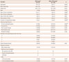
During the study period, 146 KD patients were admitted and treated with IVIG at Konkuk University Hospital. Among those, 13 patients (8.9%) had fever and cervical lymphadenopathy as the sole initial presentation (LKD group). The other 133 patients did not meet the LKD criteria. As shown in Table 1, the LKD patients were significantly older (mean: 3.9 years vs. 2.4 years) and admitted earlier (mean: 2.9 days vs. 4.6 days) than the Other-KD patients. However, the LKD patients were diagnosed and treated with IVIG significantly later (mean: 5.9 days vs. 4.9 days) than the Other-KD patients. The LKD patients were treated with antibiotics for 3 days between admission and diagnosis, while the Other-KD patients were treated with IVIG and aspirin a few hours after admission. Fever duration after IVIG treatment was not different between the two groups. However, the total durations of the fever and hospital stay were both significantly longer in the LKD patients. There were no differences in the percentage of male patients between the two groups. Of the diagnostic criteria for complete KD, cervical lymphadenopathy was significantly more frequent in the LKD patients than in the Other-KD patients (p=0.000). There were no significant differences in any other diagnostic criteria between the two groups. The frequency of complete KD was higher (92.3% vs. 65.4%) in the LKD patients, without significant differences. Although the LKD patients initially presented with only fever and cervical lymphadenopathy, the typical features of KD developed during the subsequent hospitalization period, after which complete KD was finally diagnosed in 12 patients (92.3%). Only one patient had incomplete KD and showed two principal clinical features plus CALs by echocardiography. The frequency of IVIG-resistant patients was not significantly different between the two groups (15.4% vs. 11.3%, p=0.65). Seventeen patients who were IVIG-resistant received additional rescue therapies with repeat high-dose IVIG (2 g/kg) in all. Additional IVIG rescue was successful in both patients of the LKD group; however, the five patients of the Other-KD group who were IVIG-resistant despite additional IVIG, received additional rescue therapy; of these, three patients received high-dose methylprednisolone, and two needed infliximab infusion.
Comparison of echocardiographic parameters and coronary artery lesions between groups
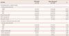
Echocardiographic parameters were compared between the two groups (Table 1). The left ventricular systolic function and the frequencies of mitral regurgitation and pericardial effusion showed no significant differences between the two groups in the acute and convalescent phase. As shown in Tables 1 and 2, in the acute phase, the incidence of CALs in the LKD patients was significantly higher than that in Other-KD patients (38.4% vs. 11.3%, p=0.02). In the acute phase, the diameters of the left main coronary artery and the anterior descending artery in the LKD patients were significantly higher than in Other-KD patients (0.95 vs. 0.63, p<0.05). However, in the convalescent phase, there were no significant differences in CALs between the two groups (15.4% vs. 4.5%, p=0.15). The diameters of the left main coronary artery, left anterior descending artery, and right coronary artery showed no statistical differences between the two groups.
Comparison of laboratory parameters and risk-scoring systems between groups
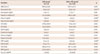
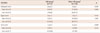
As shown in Table 3, the LKD patients had significantly elevated % neutrophil (p=0.00), CRP (p=0.01), albumin (p=0.01), and total bilirubin (p=0.02) levels. WBC count, hemoglobin, AST, and ALT were also all higher in the LKD patients, but the differences were only marginally significant (p>0.1). Platelet count, sodium, erythrocyte sedimentation rate, lactate dehydrogenase, frequency of pyuria, and BNP levels were not different between the two groups. As shown in Table 4, patient data were compared using three risk-scoring systems (Kobayashi, Egami, and Sano scores), which may be useful for predicting IVIG-resistant KD. The mean values of the Kobayashi score and the Egami score were not different between the two groups, but the mean value of the Sano score was significantly higher in the LKD group (1.23 vs. 0.68, p=0.01). However, the frequency of high-risk patients in each of the three risk scoring systems was not different between the two groups.
Discussion
Although cervical lymphadenopathy is one of the principal diagnostic criteria for KD, it is the least common, occurring in about 24% to 75% of patients with KD.6)10) Nevertheless, a few KD patients initially present with fever and cervical lymphadenopathy alone3)6)7)8)9)10)11) and are often misdiagnosed as bacterial lymphadenitis or other lymphadenopathy. Misdiagnosis can lead to unnecessary procedures, antibiotic treatment, and delay in optimal treatment.3)10) Delayed diagnosis and treatment may increase the risk of the cardiac complications associated with KD.3) Therefore, it is important to distinguish LKD patients from patients with bacterial lymphadenitis in the initial stage. Elucidating the characteristics of LKD patients may help lead to an early diagnosis and timely treatment, thereby decreasing the need for unnecessary procedures and antibiotic therapy.
April et al.10) revealed that KD patients with cervical adenopathy were significantly older (mean age: 3.4 years vs. 2.4 years) than KD patients without cervical adenopathy; in addition, these patients showed greater abnormalities in markers of systemic inflammation in the group with adenopathy, possibly due to higher inflammatory response by the more mature mucosal immune system in older children, leading to regional adenopathy in KD patients with cervical adenopathy.10) However, no differences were noted between the two groups in the prevalence of coronary artery abnormalities. Kao et al.7) examined 14 KD patients presenting with cervical lymphadenitis or a deep neck infection. The mean age of these patients was 54 months and the mean duration between diagnosis of KD and the onset of the disease was 8.2 days (6 to 20 days). Three (21.4%) patients developed CALs. Kubota et al.6) reported 29 patients initially presenting with only cervical lymphadenopathy and fever (LKD patients). Relative to KD patients with other presentations (Other-KD patients), these LKD patients were older (mean age, 4 years vs. 1 year) and admitted earlier (3 days vs. 4 days). CRP levels were significantly higher (7.6 mg/dL vs. 5.8 mg/dL) in the LKD patients than Other-KD patients. The incidence of CALs was higher (13.8% vs. 8.4%) in the LKD patients than in Other-KD patients, without significant differences. The proportion of non-responders was not different (15.4% vs. 15.5%). The results indicated that the LKD patients were older, admitted earlier, and had higher inflammatory marker levels such as CRP and % neutrophils, with some differences, which is consistent with previous reports.
First, in the present study, total bilirubin levels were significantly higher in the LKD patients, and the levels of AST and ALT were higher in the LKD patients, although the differences were only marginally significant. Eladawy et al.16) reported that patients with abnormal liver function test results were at a higher risk for IVIG resistance. However, in the present study, IVIG responsiveness was not different between the two groups (15.4% vs. 11.3%). The exact etiology of abnormalities of liver function tests in KD has not been established. Hypotheses included generalized inflammation, vasculitis, congestive heart failure secondary to myocarditis, nonsteroidal anti-inflammatory antipyretics, toxin-mediated effects, or a combination of these events.16) In order to determine whether there was a difference in IVIG responsiveness between the two groups, three risk-scoring systems, which were developed to identify children at highest risk of IVIG resistance, were applied to our data. However, the frequency of high-risk patients in each of the three risk scoring systems was not different between the two groups.
Second, in the present study, the incidence of CALs among the LKD patients was not different during the convalescence stage. Interestingly, the incidence of transient CALs was significantly higher (38.5% vs. 11.3%) in the acute stage. In the early stage of CALs (7-9 days after disease onset), an influx of neutrophils occurs in an affected coronary artery.17) This stage involves the destruction of the internal elastic lamina followed by myofibroblast proliferation, which leads to the formation of a coronary aneurysm. To prevent changes in the coronary artery, immediate treatment is necessary before pathological changes become irreversible.17)18) The mean time until diagnosis was 8.2-9.9 days in previous reports, with a high rate of CALs (21.4-70%).7)11)12)13)14)15)16)17)18)19) The mean time to diagnosis was 5.9 days in the LKD patients in the present study. Although mean duration until diagnosis may not be the only risk factor for CALs, early diagnosis before irreversible vascular changes could explain the low incidence of CALs in our study. Delayed diagnosis of KD is an apparent risk factor for CALs.19)
Whether the presence of cervical lymphadenopathy in KD is an independent risk factor for CALs is controversial. Nomura et al.9) demonstrated that patients with complete KD and only fever and cervical lymphadenopathy at admission (KDiL), showed a significantly higher frequency of IVIG resistance (38% vs. 10%) and CALs (25% vs. 4.7%). This may not be caused by delayed or early treatment, but instead by the disease severity. Compared with a previous study,6) the difference in the outcomes may indicate that patients with KDiL comprise a subset of patients with severe KD, as the incidence of patients with LKD was higher than the incidence of patients with KDiL (21% vs. 9%) among the total number of patients with KD.9) According to our results, this observation may not be true. Though the incidence of LKD patients (8.9% vs. 8.6%) and the mean time to diagnosis (5.9 days vs. 5.1 days) were very similar to the report of Nomura et al.,9) our study showed that LKD patients did not have a greater risk of CALs or IVIG resistance. Our data also showed that additional rescue therapy other than IVIG was needed only in Other-KD group, indicating that LKD is not a severe form of KD associated with increased risks of IVIG resistance and CALs.
Despite the importance of early diagnosis, it is not easy to differentiate LKD patients from patients with cervical lymphadenitis. Yanagi et al.3) reported useful cut-off values to discriminate LKD from other cervical lymphadenitis at admission including, age, 5.0 years; neutrophil counts, 10000/µ; CRP, 7.0 mg/dL; and AST, 30 IU/L. Some studies indicate that neck US and CT may be helpful for the diagnosis of KD patients with cervical lymphadenopathy at an early stage of the disease.3)20) Unfortunately, we could not compare the imaging findings between the two groups because only 9 patients in the LKD group underwent evaluation of cervical lymph nodes by US (7 patients) or CT (2 patients). The maximum diameter of cervical lymph nodes was 35 mm in CT and 25 mm in USG, respectively.
Our study has some limitations. Firstly, it is a retrospective and single-institute study and the number of patients is small. In addition, we could not completely rule out the possibility that some patients may have another disease mimicking KD, although we tried to rule out other febrile illnesses, as described previously. A multicenter study with a larger number of patients is required to elucidate the exact clinical characteristics of LKD patients and identify a useful diagnostic tool for those with early presentation.
In conclusion, the LKD patients were older, admitted earlier, and had higher inflammatory marker levels than Other-KD patients, but did not have a greater risk of IVIG resistance and CALs at the convalescent stage. These findings indicated that the KD patients with only fever and cervical lymphadenopathy as a first presentation might not be considered as a severe form of KD. Because LKD patients showed transient CALs at an early stage and delayed diagnosis is a well-known risk factor for CALs as a complication of KD, echocardiography might be helpful at an early stage of the disease to differentiate LKD patients from those with only fever and cervical lymphadenopathy who are unresponsive to empirical antibiotics.
 XML Download