

 PDF
PDF ePub
ePub Citation
 Print
Print


Introduction
Myxomas are the most common type of primary cardiac tumors, representing 30-50% of all cardiac tumors.1) Typically, myxomas manifest as a solitary, pedunculated, mobile mass in the left atrium (LA) with a stalk that is attached to the interatrial septum. Echocardiography is the most commonly used diagnostic modality. However, the echocardiographic diagnosis of myxomas is not always straightforward, and the differential diagnosis includes other benign and malignant primary heart tumors, metastatic tumors, and intracardiac thrombi. The distinction between myxomas and thrombi may pose considerable diagnostic challenges, and difficult cases have been reported.2)3) A precise diagnosis is crucial as the treatment approach and the accompanying risks differ in these two conditions. We describe a case of a LA mass that developed 1 month after surgical resection of a LA myxoma, presenting the difficulty in differential diagnosis between a recurrent myxoma and a postoperative thrombus.
Case
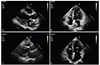
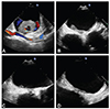
A 72-year-old Korean woman presented to our emergency department with right upper limb hemiparesis without speech impairment. Her medical history was significant for hypertension, hyperlipidemia, and diabetes. She reported no previous history of cigarette smoking and alcohol drinking. On admission to the hospital, her right upper limb hemiparesis resolved rapidly and completely. The results of laboratory examinations, including complete blood count, serum electrolyte levels, and coagulation studies, were within normal limits, except for slight hypertriglyceridemia. An electrocardiogram showed a normal sinus rhythm, and a brain computed tomography scan appeared normal. Urgent magnetic resonance imaging (MRI) of the brain revealed no definite evidence of acute infarction. Neither significant steno-occlusive lesions nor cerebral aneurysms in the intra/extracranial vessels were demonstrated by magnetic resonance angiography (MRA). Transthoracic echocardiography (TTE) showed a well-defined echogenic mass in the LA with a broad-based attachment to the interatrial septum (Fig. 1). The LA was not enlarged, and left ventricular (LV) systolic function was normal, with an ejection fraction (EF) of 60%. Transesophageal echocardiography (TEE) confirmed the presence of a heterogeneous mobile mass (33×25 mm) with internal echo-free spaces, and color Doppler flow mapping showed flow signal within the mass suggesting hypervascularity (Fig. 2A). The mass did not involve the heart valves directly, and Doppler echocardiography revealed no impairment of flow across the mitral valve. The diagnosis of an embolic transient ischemic attack (TIA) caused by a LA myxoma was made, and the ABCD2 score was 6. She was urgently referred for surgical intervention to prevent further embolic strokes.
A median sternotomy was performed and right atriotomy was created with a trans-septal approach. The LA mass was then excised en bloc for complete resection and the iatrogenic atrial septal defect was directly closed without using a patch. Histopathological examination confirmed a cardiac myxoma with a clear resection margin, and without an overlying thrombus. The postoperative course was uneventful and free of major complications. On postoperative day 4, TTE demonstrated no evidence of any remnant mass or thrombus in the LA (Fig. 1). The patient was discharged without antiplatelet therapy or anticoagulants.
One month after surgery, the patient was readmitted to our hospital because of a transient left-sided weakness in her arm and leg without speech impairment. Her neurological examination was normal, and brain MRI performed using the stroke protocol demonstrated no acute lesion with diffusion restriction. MRA of the cranial vessels showed neither steno-occlusive lesions nor aneurysmal dilatations, which was the same as before. TTE and TEE were performed to identify the source of the TIA. Scanning of the LA revealed a polypoid mass (13×7 mm) attached to the mid portion of the interatrial septum, with an irregular lobulated surface (Fig. 2B). Lipomatous hypertrophy of the interatrial septum was also noted, and no thrombus was seen in the left atrial appendage. Although LV systolic function was slightly decreased (EF 47%) without regional wall motion abnormalities, the echocardiographic findings of the mass in question were more suggestive of a remnant myxoma. However, given the short period of time after the surgery, thrombus formation at the site of direct closure of the interatrial septum was a reasonable possibility. Therefore, after discussion, intravenous heparin administration was started as a therapeutic trial and it was continued to help differentiate a remnant mass from a thrombus.
One week after the initiation of anticoagulation, a follow-up TEE examination revealed a relevant reduction in the burden of the mass in question from 13×7 mm to 8×3 mm, suggesting that the mass was most likely to be a thrombus (Fig. 2C). Therefore, long-term anticoagulation with warfarin was continued to maintain an international normalized ratio of 2-3. At the 4-month follow-up after warfarinization, the mass attached to the interatrial septum had resolved completely on TEE examination, confirming the diagnosis of a thrombus (Fig. 2D). The patient is now being maintained on warfarin and kept under regular follow-up.
Discussion
Myxomas are the most common primary cardiac tumors, although their diagnosis is not always straightforward. Most often, the typical morphological characteristics of atrial myxomas can be readily described using echocardiography, but the imaging appearance of myxomas sometimes mimics the appearance of thrombi.2) Differential diagnosis is more difficult when a recurrent mass is observed after successful surgery of a LA myxoma. Myxomas are larger, pedunculated, mobile, and most frequently attached by a stalk to the fossa ovalis of the LA septum. Less commonly, they are located in the atrial dome, mitral, pulmonic, or aortic valves, or the right atrial septum and free wall.4) On the other hand, thrombi that are generally attached to the posterior left atrial wall have a broad base, and are mostly immobile. The left atrial appendage is a useful landmark for differentiating tumor from thrombus. The formation of thrombi usually occurs in patients with organic heart disease, and in association with low cardiac output or atrial fibrillation.1) These characteristics, however, are not specific enough to always reliably distinguish left atrial thrombi from myxomas.3)
In our present case, the questionable finding was a LA mass that developed 1 month after primary surgical resection of the original tumor. It was important to differentiate a remnant myxoma from a postoperative thrombus because prompt surgical excision would have been required to prevent further embolic events in the case of a remnant tumor. Our present case had undergone uncomplicated en bloc resection. The absence of atrial fibrillation, enlarged atrial chamber, valvular heart diseases, and spontaneous atrial contrast echoes were the features weighing against the diagnosis of a thrombus. However, considering the rarity of remnants after successful surgical resection of non-familial sporadic myxomas and the rapid growth rate of the mass in question, a treatment test with anticoagulation was warranted in order to avoid an unnecessary surgical intervention.
Embolization is the second most common initial presentation of a myxoma, occurring in 30-40% of the patients. Prompt surgical excision must be performed as soon as the diagnosis is confirmed because of the high risk of new embolic complications or sudden death.1) However, postoperative thromboembolic events occur infrequently, and atrial arrhythmias are the most common complications after surgery for myxoma.5)6)7)8) Although myxomatous fragments of a tumor or thrombi at the tumor surface may dislodge during surgery, the occurrence of atrial fibrillation is the main reason for postoperative thromboembolic complications.9) Thrombus formation at the closure site is an unusual but potential complication contributing to recurrent neurologic events. However, an intracardiac thrombus after surgery for myxoma has rarely been reported, but thrombus formation after repair of an atrial septal defect or a patent foramen ovale has been reported.10)11)12) One interesting point is that all these case reports showed thrombus formation in the right atrium along the primary suture line: one case showed a pedunculated thrombus along the right atrial wall,11) whereas the other two cases showed a thrombus attached to the right atrial side of the interatrial septum.10)12) Considering low blood flow velocity in the right atrium, short-term anticoagulation has been suggested to prevent this complication after primary surgical or device closure of the atrial septal defect or a patent foramen ovale.11) Our case is unique as it showed that thrombus formation can occur in the left atrium after primary closure of the atrial septum without using an artificial atrial patch.
Removal of an atrial myxoma carries an operative mortality rate of 5% or less, and the mortality of patients waiting for surgery is 8%.1)13) Atrial thrombi misdiagnosed as myxomas can lead to an unnecessary surgical resection.3)14)15) Although a trial of anticoagulation with intravenous heparin is advised for making the differential diagnosis, it may not be applicable in case of postoperative thrombi, which can be more or less resistant to anticoagulation therapy.11) An organized thrombus associated with underlying structural heart diseases may also be less likely to respond to lytic or anticoagulation therapy.16)
In summary, the echocardiographic distinction between myxomas and thrombi is not always straightforward due to their morphological similarities. Our present case highlights 1) the importance of clinical suspicion of intracardiac thrombi after successful primary closure of the atrial septum; 2) the advantage of a clinical therapeutic trial of short-term anticoagulation with serial follow-up TEE, which could discriminate between a postoperative thrombus and a remnant cardiac tumor, resulting in avoidance of an unnecessary second-look operation. The management of an atrial mass should be based on the clinical situation (accompanying heart disease, postoperative period, etc.) and the echocardiography findings (well-limited mass, echogenicity, etc.). A treatment test with anticoagulation is worthy of attempting in cases in which there is a diagnostic challenge in differentiating myxomas from thrombi.
 XML Download