

 PDF
PDF ePub
ePub Citation
 Print
Print


Introduction
Takayasu's arteritis (TA) is a non-atherosclerotic chronic inflammatory disease of the large and medium-sized arteries resulting in obstruction or less frequently aneurysm formation. It predominantly affects young females and in general, the pathogenesis and the natural course of the disease remain poorly defined. Clinical manifestations are variable, depending on the affecting vessels and the patterns of the disease. The rarity of the disorder and heterogeneous nature of the clinical presentation predispose to late diagnosis and delayed treatment.1) We described a case of a young female patient with spontaneous resolution of the obstruction of the left subclavian artery over a 10 year interval.
Case
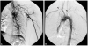
A young female underwent bilateral coronary ostioplasty at the age of 28 under the impression of TA involving both coronary ostia. Initially, she visited the hospital complaining of headache and dizziness. The symptoms had developed insidiously the previous year, and at presentation, she also felt claudication of her left arm. On physical examination, her left radial pulse was not easily palpated and carotid bruits were heard on both sides. Carotid sonography revealed diffuse wall thickening of the carotid arteries without significant obstruction of the blood flow. There were no significant abnormalities on brain imaging studies. Aortography to rule out TA revealed total occlusion of the left subclavian artery with abundant collateral formation (Fig. 1A). A segmental stenosis of the right subclavian artery and the left vertebral artery was also noted (Fig. 1B). Despite the absence of cardiac disease symptoms other than palpitation, diffuse ischemic change was seen on the electrocardiogram; hence, coronary angiography was conducted, revealing bilateral coronary ostial stenosis i.e., right 80% and left 70%. Surgical treatment of the coronary lesions following a course of steroid therapy was planned as the first priority and definitive treatment of the other vascular lesions were deferred. The initial erythrocyte sedimentation rate (ESR) was 32 mm/hr (normal range 0-20). Serum immunoglobulin G and immunoglobulin M were 1760 mg/dL (normal range 694-1618) and 188 mg/dL (normal range 60-263), respectively. Anti-DNA antibody and antinuclear antibody were negative. The patient was given oral prednisolone 50 mg a day for 7 days and subsequently tapered to 10 mg a day before surgery. The ESR fell to 7 mm/hr. On surgery, the wall of the ascending aorta around the coronary ostia appeared uniform and remarkably thickened, without calcification or atheromatous change. Both coronary ostia were successfully widened with an autologous saphenous vein patch. Her recovery was uneventful. Oral steroids at a dose of 12.5 mg a day was continued until discharge.2) Pathologic exam of the surgical specimen taken from the thickened aortic wall showed significant intimal thickening, medial fibrosis and mild chronic inflammatory infiltrates in media, which were highly consistent with TA.
Her presenting symptoms were well controlled with the medical treatment and oral steroid was tapered off over 2 months after discharge. She has since been intermittently followed-up. No specific medication was given except aspirin 100 mg a day. A 10 year post-operative follow-up coronary angiogram showed widely patent bilateral coronary ostia. Surprisingly, the obstructive lesions of the left subclavian had disappeared completely and blood flow was well maintained. The stenotic lesions of the right subclavian artery and the left vertebral artery were also resolved (Fig. 2).
Discussion
Takayasu's disease is a well-known systemic inflammatory vascular disease predominantly affecting the aorta and its major branch vessels. Systemic symptoms such as fever, malaise, night sweats or polyarthralgia may predominate at the onset of the disease. Medical treatment mainly consists of corticosteroids and immunosuppressive agents, with varying degrees of response and side effects. Surgical intervention is practiced to relieve critical obstruction, dissection or aneurysm formation but the long term outcome is often unfavorable and relapses are not uncommon.1) In a cohort of 126 TA patients, mortality was increased, as compared with the general population. Hypertension and congestive heart failure commonly complicate the disease and significantly influence the prognosis.3)
The disease is well known for its chronicity. It was previously thought to be self-limited and to follow a benign course, however, it typically relapses between long-term remissions. In fact, vascular inflammation continues to progress even in patients who appear clinically quiescent. Disease control commonly involves chronic glucocorticoid treatment, frequently leading to significant morbidity and disability.4)
Inflammatory markers are often inaccurate in determining the activity of the disease. Current vascular imaging studies, although very useful in defining vascular anatomy, also generally fail to delineate the degree of vascular inflammation that could guide the response of medical therapy.1)
Spontaneous remission is rarely reported with only 2 reports of cases exclusively affecting the renal artery.
This was the first report on spontaneous resolution of vascular obstruction at the subclavian artery.5)6) The possibility of obstruction from any other causes such as temporary vascular spasm was highly unlikely, due to the formation of abundant collaterals (Fig. 1). These collaterals had actually disappeared, as shown in Fig. 2. Aspirin is recommended as the first line medication; however the patient was on long-term aspirin therapy and its positive effect on the disease course is unclear.
It could be argued that the other chronic inflammatory vascular conditions mimicking TA were surely eliminated. Moreover, there are no pathognomic features for the definite diagnosis of TA. Laboratory parameters including numerous inflammatory markers are generally non-specific, often delayed and misleading. Nonetheless, typical clinical features of TA, as seen in this case, strongly favor the diagnosis of TA. Giant cell arteritis (GCA), which is the other main cause of large and medium vessel arteritis, can be easily differentiated from TA, since TA is typically seen in younger patients with age of onset less than 40 years and with a striking female preponderance, whereas the onset of GCA is after the age of 50 years.1) Other less commonly encountered causes of large and medium vessel arteritis, such as rheumatoid arthritis, systemic lupus erythematosus, spondyloarthopathies, Beçhet disease, sarcoidosis and polyarteritis nodosa can also be ruled out by the absence of relevant symptoms and signs.
On retrospect, the inflammatory process of the coronary ostial lesions might have been relieved by medical treatment alone and coronary surgery could have been avoided. The exact mechanism of spontaneous resolution in this case remains unclear. However, progressive vascular stenosis or occlusion does not necessarily reflect inflammatory activity of ongoing TA, but could simply indicate advancing fibrosis.1)
As shown in this case, spontaneous resolution of the vascular obstruction in TA is possible. New insights into the clinical course and outcomes across different racial and ethnic groups are necessary and the treatment strategy might be influenced by further elucidating the evolutional patterns of the disease.

 XML Download