Journal List > Korean Circ J > v.46(5) > 1017388

Jang, Shin, Bae, Jin, Seo, Yang, Kim, Cho, Kim, Park, Je, and Kim: Diagnostic Performance of Intravascular Ultrasound-Derived Minimal Lumen Area to Predict Functionally Significant Non-Left Main Coronary Artery Disease: a Meta-Analysis

Abstract

Background and Objectives

Intravascular ultrasound (IVUS)-guided percutaneous coronary intervention frequently results in unnecessary stenting due to the low positive predictive value of IVUS-derived minimal lumen area (MLA) for identification of functionally significant coronary stenosis. We appraised the diagnostic accuracy of IVUS-derived MLA compared with the fractional flow reserve (FFR) to assess intermediate coronary stenosis.

Subjects and Methods

We searched MEDLINE and Cochrane databases for studies using IVUS and FFR methods to establish the best MLA cut-off values to predict significant non-left main coronary artery stenosis. Summary estimates were obtained using a random-effects model.

Results

The 17 studies used in our analysis enrolled 3920 patients with 4267 lesions. The weighted overall mean MLA cut-off value was 2.58 mm2. The pooled MLA sensitivity that predicted functionally significant coronary stenosis was 0.75 (confidence interval [CI]: 0.72 to 0.77) and the specificity was 0.66 (CI: 0.64 to 0.68). The positive likelihood ratio (LR) was 2.33 (CI: 2.06 to 2.63) and LR (-) was 0.33 (CI: 0.26 to 0.42). The pooled diagnostic odds ratio (DOR) was 7.53 (CI: 5.26 to 10.76) and the area under the summary receiver operating characteristic curve for all the trials was 0.782 with a Q point of 0.720. Meta-regression analysis demonstrated that an FFR cut-off point of 0.75 was associated with a four times higher diagnostic accuracy compared to that of 0.80 (relative DOR: 3.92; 95% CI: 1.25 to 12.34).

Conclusion

IVUS-derived MLA has limited diagnostic accuracy and needs careful interpretation to correlate with functionally significant non-left main coronary artery stenosis.

Introduction

Fractional flow reserve (FFR) is an invasive physiologic assessment of significant ischemia and is an important tool to determine whether to proceed with percutaneous coronary intervention (PCI) of intermediate coronary stenosis.1) Moreover, PCI of coronary stenosis with FFR is greater than either 0.75 or 0.80 without intervention has been safe and cost-effective.2)
On the other hand, intravascular ultrasound (IVUS) has been widely used to assess coronary stenosis, either quantitatively or qualitatively. IVUS has been reported to improve clinical outcomes compared to PCI guided by angiography alone.3) Meanwhile, IVUS-derived minimal lumen area (MLA) has been proposed as a simple anatomic alternative to FFR to determine the severity of intermediate coronary stenosis.4) Although MLA <4.0 mm2 has been widely used to predict the functional significance of stenosis in non-left main coronary artery,4)5) reported IVUS-derived MLA cutoff threshold ranges from 2.0 to 4.0 mm2 and the use of IVUS to guide PCI, has resulted in unnecessary stenting approximately half of the time because of a relatively low positive predictive value.6)
In order to elucidate whether MLA derived from IVUS can be used as an indicator in the diagnosis of functionally significant coronary artery stenosis, we conducted a systematic review and meta-analysis of studies evaluating the diagnostic accuracy of IVUS-derived MLA for the assessment of intermediate coronary lesions.

Subjects and Methods

Data sources and searches

We identified relevant studies through electronic searches of PubMed and Ovid MEDLINE and the Cochrane Central Register of Controlled Trials through January 2015. A systematic search was performed with the Medical Subject Headings terms and title/abstract words: ("Ultrasound" [Mesh] OR "Ultrasound [Title/Abstract] OR "Ultrasonography [Mesh]" OR "Ultrasonography" [Title/Abstract] OR "Ultrasonics" [Mesh] OR "Ultrasonics" [Title/Abstract]) AND ("Fractional" [Title/Abstract] AND "Flow" [Title/Abstract] AND "Reserve" [Title/Abstract]).

Study selection

Two investigators (J.-S.J. and H.-Y.J.) independently inspected the title and abstract of each citation to identify those studies reporting the diagnostic value of IVUS-derived MLA and then obtained the full text. Inclusion criteria for the primary studies were as follows: 1) studies which measured IVUS and FFR in the same set of patients at the time of coronary angiography or PCI 2) studies providing diagnostic performance test data and 3) establishment of best cut-off value for MLA based on diagnostic tests.

Quality assessment

Two reviewers (J.-S.J. and H.-Y.J.) used 11 items from the published quality assessment for studies of diagnostic accuracy (QUADAS) guidelines recommended by The Cochrane Collaboration Methods group, regarding screening and diagnostic tests as a tool to assess the included studies (Supplementary Fig. 1 in the online-only Data Supplement).7) Disarrangements were resolved by consensus.

Data synthesis and analysis

A random effects model was used to differentiate the predictive accuracy of IVUS-MLA between different studies because of variability between studies. We applied likelihood ratios (LR) for alternative statistics to sensitivity and specificity in summarizing the properties of a prognostic test.8) In the context of our analysis, the LR describes how many times likely patients who have a functionally significant CAD have that result than patients without a significant CAD. Because cut-off points of FFR for defining a functionally significant CAD were different across studies, we calculated the Spearman's correlation coefficient between sensitivity and specificity. We used diagnostic odds ratio (DOR) to describe the odds of positive test results in participants with functionally significant disease compared with the odds of positive test results in those without significant disease.
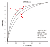
Additionally, sensitivities and specificities were summarized using a receiver operating characteristic (ROC) curve, where the diagnostic accuracy is shown by plotting 1-specificity against specificity. To summarize the curve, the area under the curve and the Q point (Q*) index were used. The point of Q (Q*, sensitivity=specificity) obtained from the summary ROC curve was used as a measure of global accuracy.9)
A chi square test was used to detect statistically significant heterogeneity. Statistical heterogeneity across studies was quantified with the Cochran's Q and I2 statistic, which is derived from Cochran's Q and the degrees of freedom [I2=100%×(Cochran Q –degrees of freedom)/Cochran Q].10) We conducted subgroup- and meta-regression analyses to detect the heterogeneity between studies. To evaluate the statistical outcome variability, we detected the pooled outcomes by sensitivity analyses.
All statistical analyses were performed using the Review Manager version 5.1 (The Nordic Cochrane Center, Copenhagen, Denmark) and Meta DiSc version 1.4 (Romany Cajal Hospital, Madrid, Spain) programs.

Results

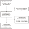
A total of 331 publications were reviewed and 28 studies were selected for inclusion and further evaluated. Three studies were excluded because the left main coronary artery lesions were primarily evaluated. These left main studies suggest that MLA values from 4.1 to 5.9 mm2 can predict functional significance of left main disease with an FFR cut point of 0.75 or 0.80.11)12)13) Seventeen non-left main clinical studies were subsequently included into the final analysis (Fig. 1).
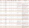
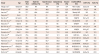
Characteristics of the included studies are summarized in Table 1. A total of 3920 patients and 4267 lesions were included in the analysis. Three studies were retrospective observational studies14)15)16) and 14 studies were prospective cohort studies with consecutive patients.4)5)6)17)18)19)20)21)22)23)24)25)26)27) Ten studies were done in patients with Asian ethnicity,4)6)14)16)17)18)22)23)25)27) whereas six studies were performed in the Western patient population.5)15)19)20)21)24) One study enrolled patients from both regional groups.26) The FFR cut-off values defining functionally significant coronary stenosis were <0.75 in three studies4)5)17) and <0.80 in 13 studies.6)14)15)16)18)20)21)22)23)24)25)26)27) One study used both FFR cut-off values for different IVUS-derived MLA.19) The weighted overall mean MLA cut-off value was 2.58 mm2 ranging from 2.00 to 4.00 mm2. To compare possible differences between studies, the prevalence of risk factors (diabetes, hypertension, hyperlipidemia, current smoking), distribution of involved coronary arteries, left ventricular ejection fraction, and proportion of patients with acute coronary syndrome were extracted (Table 2).
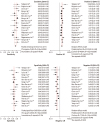
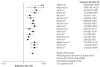
When all the trials were pooled, the pooled sensitivity of MLA predicting functionally significant coronary stenosis was 0.75 (confidence interval [CI]: 0.72 to 0.77; I2=78.9%) and specificity was 0.66 (CI: 0.64 to 0.68; I2=67.4%). The positive LR was 2.33 (CI: 2.06 to 2.63; I2=67.2%) and LR (-) was 0.33 (CI: 0.26 to 0.42; I2=79.4) (Fig. 2). The pooled DOR was 7.53 (CI: 5.26 to 10.76; I2=77.6; Fig. 3) and the area under the summary ROC curve for all the trials was 0.782 with the Q* of 0.720 (Fig. 4).

Sub-group and meta-regression analyses

Stratified analysis according to different FFR cut-off values revealed a trend of better diagnostic performance with trials using an FFR cut-off of 0.75 compared with trials using an FFR value of 0.80 (DOR: 21.61; CI: 10.59 to 44.11 vs. DOR: 6.59; CI: 4.62 to 9.39).
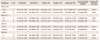
Subgroup analysis of trials performed in Asian populations revealed a DOR of 8.46 (CI: 5.92 to 12.10), whereas that of trials in Western populations was 5.37 (CI: 3.49 to 8.27). In addition, diagnostic performance of IVUS-MLA was increased in studies with QUADAS scores ≥7 (DOR: 8.76; CI 5.27 to 14.56) compared to studies with QUADAS scores <7 (DOR: 6.80; CI: 4.24 to 10.92) (Table 3).
Possible sources of heterogeneity across the studies were explored using meta-regression analysis with five covariates as predictor variables: FFR cut-off values, MLA cut-off values, QUADAS score, study design, and ethnicity of the involved patients. An FFR cut-off of 0.75 was associated with four times higher diagnostic accuracy compared to the value of 0.80 (relative DOR: 3.92; 95% CI: 1.25 to 12.34; p=0.023).

Discussion

In the present meta-analysis of 17 observational studies, consisting of 3920 patients, we found that IVUS-derived MLA has a limited but acceptable pooled diagnostic performance in predicting functionally significant non-left main coronary artery disease with a pooled DOR of 7.53 and the area under the summary ROC curve of 0.782. Furthermore, we found that the diagnostic performance of IVUS-derived MLA was higher in Asian population studies when compared to studies performed in Western populations. We also found that an FFR cut-off of 0.75 is associated with better diagnostic accuracy compared to an FFR cut-off of 0.80.
Despite promising data about the use of anatomical variables to correlate the functional significance of coronary stenosis, most physicians do not believe that anatomical predictors can substitute for FFR. Even though IVUS has excellent spatial resolution and provides information to guide intervention, lumen assessment alone does not reflect microcirculatory function, collateral circulation, and viability of the corresponding myocardium.28) Nevertheless, many physicians use IVUS to guide interventional treatment in lieu of FFR for several reasons. The FFR test requires additional equipment and expert personnel to measure and interpret the results. Contrary to FFR, IVUS is relatively simple to perform and its use is associated with low complication rates. In addition, the operators do not need to use intracoronary adenosine to induce maximal hyperemia, which puts the patient at risk. Although the diagnostic accuracy of IVUS is not enough to substitute for FFR, both IVUS and FFR may be used as complementary tools in the catheterization laboratory to provide both functional and anatomical data to guide optimal decision-making in patients with intermediate coronary stenosis.
Previous meta-analysis comparing IVUS-MLA versus FFR for the assessment of intermediate lesions revealed limited accuracy of IVUS imaging in non-left main lesions to predict functionally significant stenosis as compared with FFR, while better accuracy was shown in left main lesions.29) The results of our study support data generated by a previous study. However, we included and analyzed data from several other recently published studies.15)16)22)23)24)25)26)27) We did not include studies performed exclusively on left main disease subsets because we thought that left main stenosis has a different clinical meaning with different IVUS-MLA cut-offs compared with other vessels. Therefore, left main disease needs to be assessed separately. In addition, we performed several subgroup analyses according to FFR cut-off values, MLA, ethnicity, and QUADAS scores of the included studies to better understand and confirm benefits in certain subgroups compared with others.
Contrary to the earlier reports using an FFR cut-off value of 0.75 to define functional significance of coronary narrowing.4)5)17) Recent studies adopted a less restrictive and more clinically relevant cutoff value of 0.80 to minimize the number of untreated lesions that may cause significant ischemia.6)18)30) Even though subgroup- and meta-regression analysis of our study suggests greater diagnostic performance of IVUS-derived MLA in studies using an FFR cut-off point of 0.75 compared to studies using 0.80, it is not appropriate to adopt 0.75 as the standard cut-off point for functionally significant coronary disease. This is due to routine measurements of FFR and stenting limited to stenoses with an FFR ≤0.8, which significantly lowered the rate of the composite endpoint of death, myocardial infarction, and repeat revascularization at one year compared with stenting of all of the lesions deemed appropriate based on the angiography.30)
In our analysis, mean MLA cut-off in Asian populations was 2.68 mm2 and that of Westerners was 3.03 mm2. The difference in mean MLA cut-off value by ethnicity is one of the interesting and novel findings in our study. Han et al.26) found different demographic and lesion characteristics, as well as, different cut-off values between Asians and Westerners. We postulated that lower body mass index and smaller myocardial masses in Asian people may result in lower MLA cut-off values and different physiologic significance of coronary stenosis when compared with Western populations.
There are several limitations to be addressed in our study. First, the majority of studies included in our analysis were observational studies from different cohorts with no randomized controlled trials. This caused our results to have insufficient power. Second, the proportion of the involved coronary arteries and extent of coronary diseases were different across the included studies. It was not possible to suggest the diagnostic performance of IVUS-MLA according to the lesion location. Further, we could not perform separate subgroup analyses of all coronary arteries and their location (proximal-, mid-, distal-) because very few studies presented such data. Moreover, we could not differentiate patients presenting with stable angina and acute coronary syndrome, despite differences in clinical significance of IVUS-derived MLA and FFR. Additionally, the IVUS criteria to discriminate the functional significance of lesions in different locations were applied differently across studies. Last, we did not take into account the plaque composition that can affect clinical outcomes.
In conclusion, IVUS-derived MLA has limited diagnostic accuracy in predicting functionally significant coronary artery disease and cannot be used alone to make the decision whether or not to proceed with the PCI of intermediate non-left main coronary artery stenosis.

Figures and Tables

Fig. 1

Trial flow chart shows number of studies retrieved by individual searches and number of trials included in review.

kcj-46-622-g001
Fig. 2

Combined sensitivity, specificity, positive and negative LRs of the included trials. The MLA derived from IVUS has a limited pooled diagnostic performance in predicting functionally significant non-left main coronary artery stenosis. LR: likelihood ratio, MLA: minimal lumen area, IVUS: intravascular ultrasound, CI: confidence interval.

kcj-46-622-g002
Fig. 3

Pooled DOR of the included studies. The odds of positive intravascular ultrasound results were 7.53 times higher in patients with functionally significant disease compared to the odds of positive results in patients without significant disease. DOR: diagnostic odds ratio.

kcj-46-622-g003
Fig. 4

Summary ROC curve of the included studies. The area under the summary ROC curve for all the trials was 0.782 with the Q point (Q*) of 0.720. The upper and lower lines indicate 95% confidence intervals. ROC: receiver operating characteristic. SROC: summary receiver operating characteristic, AUC: area under the curve, SE: standard error.

kcj-46-622-g004
Table 1

Characteristics of included studies

kcj-46-622-i001
Study Year Design QUADAS Ethnicity Patient/Lesion FFR cut-off MLA cut-off Inclusion criteria Exclusion criteria
Takagi et al.4) 1999 Prospective, single center 6 Asian 42/51 0.75 3.00 NA NA
Briguori et al.5) 2001 Prospective, single center 9 Western 43/53 0.75 4.0 40% to 70% diameter stenosis NA
Lee et al.17) 2010 Prospective, multicenter 8 Asian 94/94 0.75 2.0 30% to 75% diameter stenosis in a small native coronary artery (RVD <3.0 mm) Unstable ACS, fail to consent, heart rate<50 beats/min, MI, previous PCI, ostial lesion, thrombus, left main
Kang et al.6) 2011 Prospective, single center 7 Asian 201/236 0.80 2.4 30% to 75% diameter stenosis Multiple stenoses, bypass graft, left main, side branch lesions, ISR, previous PCI, MI, thrombi
Koo et al.18) 2011 Prospective, multicenter 7 Asian 252/267 0.80 2.75 30% to 70% diameter stenosis at proximal or mid part of 3 major epicardial coronary arteries STEMI, RWMAs, additional stenosis, LVEF <40%, primary myocardial or valvular disease, contraindication to adenosine, left main, collateral vessels, or thrombus
Ben-Dor et al.19) 2011 Prospective, single center 5 Western 84/92 0.80/0.75 3.2/2.82 40-70% diameter stenosis AMI, left main, saphenous vein graft, <2.5 mm in diameter, or >1 lesion
Kang et al.14) 2012 Retrospective, single center 5 Asian 692/784 0.80 2.40 30% to 90% diameter stenosis Multiple stenoses, bypass graft, left main, side branch, ISR, previous PCI, AMI, TIMI grade <3 flow thrombi
Ben-Dor et al.20) 2012 Prospective, single center 6 Western 185/205 0.80 3.09 40% to 70% diameter stenosis AMI, saphenous vein graft, <2.5 mm in diameter or >1 lesion
Kwan et al.22) 2012 Prospective, multicenter 7 Asian 169/169 0.80 3.03 40% to 100% diameter stenosis, TIMI 3 flow Multiple lesions, left main, ostial lesion, ISR, thrombus, CTO, calcified lesion, previous PCI or CABG, MI, LVEF <40%, collateral circulation, valvular or pericardial disease, pacemaker, intolerance to adenosine
Gonzalo et al.21) 2012 Prospective, single center 6 Western 56/61 0.80 2.36 40% to 70% diameter stenosis ACS, serial stenoses, or diffuse disease, previous infarction, left main, graft stenosis, contraindications to adenosine, hemodynamic instability, renal insufficiency, vessel tortuosity, severe calcification
Chen et al.25) 2013 Prospective, multicenter 6 Asian 323/323 0.80 2.97 ≥40% diameter stenosis, TIMI flow 3 Multiple lesions, left main, ostial lesion, ISR, thrombus, previous PCI or CABG, MI, LVEF<40%, collateral circulation, valvular or pericardial disease, pacemaker, intolerance to adenosine
Cui et al.23) 2013 Prospective, single center 7 Asian 141/165 0.80 3.15 40% to 70% diameter stenosis Left main, RVD <2.5 mm, multiple stenosis, bypass graft lesions, AMI, TIMI grade <3 flow, thrombi
Waksman et al.24) 2013 Prospective, multicenter 5 Western 350/367 0.80 3.07 40% to 80% diameter stenosis MI, saphenous vein graft lesions, RVD <2.5 mm, >1 lesion
Han et al.26) 2014 Prospective, multicenter 5 Asian and Western 822/881 0.80 2.75 Intermediate de novo coronary stenosis (RVD: 2.0-4.5 mm and lesion length: <40 mm) Incomplete clinical information, age <18 years, LVEF <35%, incomplete angiographic data
Naganuma et al.15) 2014 Retrospective, multicenter 5 Western 109/132 0.80 2.70 40% to 70% diameter stenosis Multiple stenoses, previous MI, left main, AMI, bypass graft disease, CTO
Yang et al.16) 2014 Retrospective, single center 6 Asian 206/206 0.80 3.2/2.5 40% to 70% diameter stenosis at the proximal or mid portion of the LAD and mean RVD ≥3.0 mm MI, LVEF<40%, RWMA, primary myocardial or valvular heart disease, additional stenosis, ISR, left main, collateral vessel and contraindication to adenosine
Doh et al.27) 2014 Prospective, multicenter 7 Asian 151/181 0.80 2.82 30% to 70% diameter stenosis ACS with RWMA, thrombus, additional stenosis, lesion length >40 mm, RVD <2.5 and >4.0 mm, LVEF<40%, primary myocardial or valvular heart disease, left main, collateral vessels, and non-diagnostic CCTA

QUADAS: quality assessment for studies of diagnostic accuracy, FFR: fractional flow reserve, MLA: minimal lumen area, NA: not applicable, ACS: acute coronary syndrome, MI: myocardial infarction, RVD: reference vessel diameter, PCI: percutaneous coronary intervention, STEMI: ST-elevation myocardial infarction, RWMA: regional wall motion abnormality, LVEF: left ventricular ejection fraction, AMI: acute myocardial infarction, ISR: in stent restenosis, TIMI: thrombolysis in myocardial infarction, CTO: chronic total occlusion, CABG: coronary artery bypass grafting, LAD: left anterior descending artery, CCTA: coronary computed tomographic angiography

Table 2

Characteristics of included patients

kcj-46-622-i002
Study Age Male (%) Diabetes mellitus (%) Hypertension (%) Dyslipidemia (%) Smoker (%) LAD/LCX/RCA (%) LVEF (%) ACS (%)
Takagi et al.4) 60.0±10.4 88.0 NA NA NA NA 49/11.7/39.2 55.5±7.8 NA
Briguori et al.5) NP 86.1 11.6 48.8 58 32.5 60.4/9.4/26.4 60±8 NA
Lee et al.17) 58 77.7 40.4 64.5 83.0 36.2 Mostly LAD NA 21.3
Kang et al.6) 61±9 72 30 61 68 49 67/11/22 61±6 28
Koo et al.18) 62.1±9.4 65 31 60 43 19.4 74/8/18 64.1±7.5 23
Ben-Dor et al.19) 63.9±11.8 58.3 NA NA NA NA 66.3/NA/NA NA NA
Kang et al.14) 62±10 72 32 59 65 49 67/9/24 61±6 22
Ben-Dor et al.20) 64.5±11.5 66.4 25.4 82.1 80.5 21 56.1/15.1/22.9 54.7±12.7 NA
Kwan et al.22) 63.2±10.4 74.6 21.9 66.9 13.6 41.4 100/0/0 60.2±8.7 65.7
Gonzalo et al.21) 62±11 83.9 33.9 71.4 73.2 44.6 49.2/24.6/26.2 NA 12.5
Chen et al.25) 63.5±10.1 75.5 26.3 68.7 12.1 40.5 LAD 52.5 58.9±7.5 21.3
Cui et al.23) 27-83 NA 35.8 68.5 56.4 31.5 67.3/13.9/13.3 66.8±8.0 NA
Waksman et al.24) 61.5±10.9 74.3 30.6 85.7 89.3 29.2 57.2/18.0/23.7 NA 42.2
Han et al.26) 63.2±10.3 70.6 33.9 70.3 65.8 NA 65.4/NA/NA 60.9±9.1 23.2
Naganuma et al.15) 68.0±9.3 89.0 24.8 52.3 58.7 11.9 50.8/28.0.21.2 55.6±8.3 9.2
Yang et al.16) 61±10 49.0 30.6 51.9 50.5 NA 100/0/0 NA 42.2
Doh et al.27) 63.2±9.6 71.5 34.7 76.2 68.2 NA 59.7/NA/NA 64.9±7.6 27.8

LAD: left anterior descending artery, LCX: left circumflex artery, RCA: right coronary artery, LVEF: left ventricular ejection fraction, ACS: acute coronary syndrome, NA: not applicable

Table 3

Subgroup analyses of IVUS-derived MLA based on various conditions

kcj-46-622-i003
Subgroups Trials Sensitivity Specificity Positive LR Negative LR Diagnostic OR Summary ROC AUC/Q* Diagnostic accuracy
FFR cut-off
 <0.75 4 0.84 (0.75-0.91) 0.77 (0.70-0.82) 3.80 (2.06-7.00) 0.21 (0.13-0.33) 21.61 (10.59-44.11) 0.8977/0.8287 0.790
 <0.80 14 0.74 (0.72-0.76) 0.65 (0.64-0.67) 2.25 (2.00-2.53) 0.36 (0.28-0.45) 6.59 (4.62-9.39) 0.7496/0.6931 0.684
MLA (mm2)
 <0.3 11 0.75 (0.72-0.77) 0.66 (0.64-0.68) 2.48 (2.10-2.92) 0.32 (0.24-0.44) 8.26 (5.16-13.22) 0.7996/0.7355 0.688
 ≥0.3 8 0.74 (0.70-0.79) 0.69 (0.65-0.72) 2.43 (1.93-3.07) 0.35 (0.26-0.48) 7.69 (4.48-13.19) 0.8026/0.7381 0.707
Ethnicity
 Asian 11 0.78 (0.76-0.81) 0.66 (0.63-0.68) 2.35 (2.09-2.64) 0.30 (0.23-0.39) 8.46 (5.92-12.10) 0.8046/0.7399 0.704
 Western 7 0.69 (0.64-0.74) 0.69 (0.66-0.73) 2.29 (1.87-2.82) 0.46 (0.38-0.57) 5.37 (3.49-8.27) 0.7580/0.7001 0.669
QUADAS
 <7 10 0.73 (0.70-0.76) 0.66 (0.64-0.68) 2.30 (1.94-2.72) 0.36 (0.26-0.49) 6.80 (4.24-10.92) 0.7808/0.7193 0.684
 ≥7 7 0.79 (0.74-0.83) 0.66 (0.62-0.69) 2.39 (2.00-2.86) 0.30 (0.22-0.42) 8.76 (5.27-14.56) 0.7938/0.7304 0.700

IVUS: intravascular ultrasound, MLA: minimal lumen area, LR: likelihood ratio, OR: odds ratio, ROC: receiver operating characteristic, AUC: area under the curve, Q*: Q point, FFR: fractional flow reserve, QUADAS: quality assessment for studies of diagnostic accuracy

Acknowledgments

This work was supported by 2015 Inje University Busan Paik Hospital research grant.

Notes

The authors have no financial conflicts of interest.

References

1. Bech GJ, De Bruyne B, Pijls NH, et al. Fractional flow reserve to determine the appropriateness of angioplasty in moderate coronary stenosis: a randomized trial. Circulation. 2001; 103:2928–2934.
2. Fearon WF, Bornschein B, Tonino PA, et al. Economic evaluation of fractional flow reserve-guided percutaneous coronary intervention in patients with multivessel disease. Circulation. 2010; 122:2545–2550.
3. Jang JS, Song YJ, Kang W, et al. Intravascular ultrasound-guided implantation of drug-eluting stents to improve outcome: a meta-analysis. JACC Cardiovasc Interv. 2014; 7:233–243.
4. Takagi A, Tsurumi Y, Ishii Y, Suzuki K, Kawana M, Kasanuki H. Clinical potential of intravascular ultrasound for physiological assessment of coronary stenosis: relationship between quantitative ultrasound tomography and pressure-derived fractional flow reserve. Circulation. 1999; 100:250–255.
5. Briguori C, Anzuini A, Airoldi F, et al. Intravascular ultrasound criteria for the assessment of the functional significance of intermediate coronary artery stenoses and comparison with fractional flow reserve. Am J Cardiol. 2001; 87:136–141.
6. Kang SJ, Lee JY, Ahn JM, et al. Validation of intravascular ultrasound-derived parameters with fractional flow reserve for assessment of coronary stenosis severity. Circ Cardiovasc Interv. 2011; 4:65–71.
7. Leeflang MM, Deeks JJ, Gatsonis C, Bossuyt PM. Cochrane Diagnostic Test Accuracy Working Group. Systematic reviews of diagnostic test accuracy. Ann Intern Med. 2008; 149:889–897.
8. Deeks JJ, Altman DG. Diagnostic tests 4: likelihood ratios. BMJ. 2004; 329:168–169.
9. Moses LE, Shapiro D, Littenberg B. Combining independent studies of a diagnostic test into a summary ROC curve: data-analytic approaches and some additional considerations. Stat Med. 1993; 12:1293–1316.
10. Dinnes J, Deeks J, Kirby J, Roderick P. A methodological review of how heterogeneity has been examined in systematic reviews of diagnostic test accuracy. Health Technol Assess. 2005; 9:1–113. iii
11. Jasti V, Ivan E, Yalamanchili V, Wongpraparut N, Leesar MA. Correlations between fractional flow reserve and intravascular ultrasound in patients with an ambiguous left main coronary artery stenosis. Circulation. 2004; 110:2831–2836.
12. Kang SJ, Lee JY, Ahn JM, et al. Intravascular ultrasound-derived predictors for fractional flow reserve in intermediate left main disease. JACC Cardiovasc Interv. 2011; 4:1168–1174.
13. Park SJ, Ahn JM, Kang SJ, et al. Intravascular ultrasound-derived minimal lumen area criteria for functionally significant left main coronary artery stenosis. JACC Cardiovasc Interv. 2014; 7:868–874.
14. Kang SJ, Ahn JM, Song H, et al. Usefulness of minimal luminal coronary area determined by intravascular ultrasound to predict functional significance in stable and unstable angina pectoris. Am J Cardiol. 2012; 109:947–953.
15. Naganuma T, Latib A, Costopoulos C, et al. The role of intravascular ultrasound and quantitative angiography in the functional assessment of intermediate coronary lesions: correlation with fractional flow reserve. Cardiovasc Revasc Med. 2014; 15:3–7.
16. Yang HM, Tahk SJ, Lim HS, et al. Relationship between intravascular ultrasound parameters and fractional flow reserve in intermediate coronary artery stenosis of left anterior descending artery: intravascular ultrasound volumetric analysis. Catheter Cardiovasc Interv. 2014; 83:386–394.
17. Lee CH, Tai BC, Soon CY, et al. New set of intravascular ultrasound-derived anatomic criteria for defining functionally significant stenoses in small coronary arteries (results from Intravascular Ultrasound Diagnostic Evaluation of Atherosclerosis in Singapore [IDEAS] study). Am J Cardiol. 2010; 105:1378–1384.
18. Koo BK, Yang HM, Doh JH, et al. Optimal intravascular ultrasound criteria and their accuracy for defining the functional significance of intermediate coronary stenoses of different locations. JACC Cardiovasc Interv. 2011; 4:803–811.
19. Ben-Dor I, Torguson R, Gaglia MA Jr, et al. Correlation between fractional flow reserve and intravascular ultrasound lumen area in intermediate coronary artery stenosis. EuroIntervention. 2011; 7:225–233.
20. Ben-Dor I, Torguson R, Deksissa T, et al. Intravascular ultrasound lumen area parameters for assessment of physiological ischemia by fractional flow reserve in intermediate coronary artery stenosis. Cardiovasc Revasc Med. 2012; 13:177–182.
21. Gonzalo N, Escaned J, Alfonso F, et al. Morphometric assessment of coronary stenosis relevance with optical coherence tomography: a comparison with fractional flow reserve and intravascular ultrasound. J Am Coll Cardiol. 2012; 59:1080–1089.
22. Kwan TW, Yang S, Xu B, et al. Optimized quantitative angiographic and intravascular ultrasound parameters predicting the functional significance of single de novo lesions in the left anterior descending artery. Chin Med J (Engl). 2012; 125:4249–4253.
23. Cui M, Zhu D, Guo LJ, et al. Usefulness of lumen area parameters determined by intravascular ultrasound to predict functional significance of intermediate coronary artery stenosis. Chin Med J (Engl). 2013; 126:1606–1611.
24. Waksman R, Legutko J, Singh J, et al. FIRST: Fractional flow reserve and intravascular ultrasound relationship study. J Am Coll Cardiol. 2013; 61:917–923.
25. Chen SL, Xu B, Chen JB, et al. Diagnostic accuracy of quantitative angiographic and intravascular ultrasound parameters predicting the functional significance of single de novo lesions. Int J Cardiol. 2013; 168:1364–1369.
26. Han JK, Koo BK, Park KW, et al. Optimal intravascular ultrasound criteria for defining the functional significance of intermediate coronary stenosis: an international multicenter study. Cardiology. 2014; 127:256–262.
27. Doh JH, Koo BK, Nam CW, et al. Diagnostic value of coronary CT angiography in comparison with invasive coronary angiography and intravascular ultrasound in patients with intermediate coronary artery stenosis: results from the prospective multicentre FIGURE-OUT (Functional Imaging criteria for GUiding REview of invasive coronary angiOgraphy, intravascular Ultrasound, and coronary computed Tomographic angiography) study. Eur Heart J Cardiovasc Imaging. 2014; 15:870–877.
28. Pijls NH, De Bruyne B, Peels K, et al. Measurement of fractional flow reserve to assess the functional severity of coronary-artery stenoses. N Engl J Med. 1996; 334:1703–1708.
29. Nascimento BR, de Sousa MR, Koo BK, et al. Diagnostic accuracy of intravascular ultrasound-derived minimal lumen area compared with fractional flow reserve--meta-analysis: pooled accuracy of IVUS luminal area versus FFR. Catheter Cardiovasc Interv. 2014; 84:377–385.
30. Tonino PA, De Bruyne B, Pijls NH, et al. Fractional flow reserve versus angiography for guiding percutaneous coronary intervention. N Engl J Med. 2009; 360:213–224.

Supplementary Materials

The online-only Data Supplement is available with this article at http://dx.doi.org/10.4070/kcj.2016.46.5.622.

Supplementary Fig. 1

Overall quality rating according to the QUADAS list. QUADAS: quality assessment for studies of diagnostic accuracy.
TOOLS
Similar articles