

 PDF
PDF ePub
ePub Citation
 Print
Print


Introduction
Atherosclerosis is a major mechanism of coronary artery disease (CAD). Although statins are currently considered the mainstay of CAD prevention, marine-derived n-3 polyunsaturated fatty acids (ω-3 PUFA), mainly composed of eicosapentaenoic acid (EPA) and docosahexaenoic acid (DHA), have received attention over the last two decades for their antiatherosclerotic effects. These effects appear to be mediated by antithrombogenic or triglyceride-lowering activities, alteration or interference in adhesion molecule metabolism, and the suppression of inflammatory mediators.1)2)3) Another study has shown a relationship between a lower ω-3 PUFA serum content and the presence of lipid-rich atherosclerotic plaques using integrated backscatter intravascular ultrasound (IVUS).4) Moreover, Nozue et al. reported that decreased ω-3 PUFA levels are associated with atheroma progression, despite low levels of low-density lipoprotein (LDL) cholesterol.5) However, recent clinical trials failed to show that ω-3 PUFA had significant protective cardiovascular benefits.6)7) These conflicting results may be explained by the fact that ω-3 PUFA shows little effect in the presence of an aggressive medical treatment background, including statins (the first-line option for primary and, especially, secondary prevention in patients with CAD).8) In this study, we examined whether ω-3 PUFA combined with statins results in additive effects that may potentially lead to a reduction of the atherosclerotic burden in CAD patients requiring coronary stent implantation.
Subjects and Methods
Study design and population
This study was a prospective randomized placebo-controlled trial conducted between September 2010 and May 2012. A total of 74 CAD patients requiring percutaneous coronary intervention (PCI) with stent implantation were enrolled (38 patients in the n-3 group and 36 patients in the placebo group). Patients underwent IVUS examinations before and after stent implantation. The exclusion criteria were as follows: no atherosclerotic plaque detected by IVUS, target lesion containing significantly big branches or calcification with a significant echo-drop out behind, age <18 years or >80 years, acute myocardial infarction (MI) within the last 72 hours, left main coronary artery disease, severe liver or renal dysfunction, left ventricular ejection fraction (LVEF)<40%, uncontrolled hypertension or diabetes, bleeding tendency, or unwilling participants. All eligible patients were prescribed statins according to clinician preference, and then randomly assigned to two groups: n-3 group (3 g of ω-3 PUFA containing 1395 mg of EPA and 1125 mg of DHA per day, n=38) and placebo group (placebo, n=36). All patients showed good compliance to their medications and completed a 12-month clinical follow-up. Blood samples were collected on the day of PCI and at the 12-month follow-up visit, and evaluated for total cholesterol, high-density lipoprotein (HDL) cholesterol, LDL cholesterol, and triglyceride levels. This study was approved by the Institutional Review Board of Pusan National University Hospital, and informed consent was obtained from all patients prior to their enrolment.
Randomization
Simple randomization was carried out using random number tables to assign each participant to the intervention or control group. Participants were assigned randomization numbers sequentially on recruitment to the study, and the randomization codes were retained by the clinical research coordinator of the present study. The personnel responsible for randomization as well as those performing laboratory measurements were blinded to the randomization assignments.
Coronary angiography and quantitative coronary angio graphic analysis
Coronary angiography (CAG) was performed using the standard radial or femoral approaches. Before the procedure, all patients received a loading dose of 300 mg of aspirin, 600 mg of clopidogrel, and 100 U/kg of heparin. Additionally, intravenous heparin was administered to maintain the activated clotting time within 250-300s. Quantitative measurements of coronary arteries were obtained using a computer-based image-analysis system with best-shown segments. The reference diameter and minimal lesion diameter (MLD) were measured offline, and percentage diameter stenosis was calculated from these values. Late loss was defined as the difference between the MLD immediately post-PCI and the MLD at the 12-month follow-up.
Intravascular ultrasound imaging and analysis
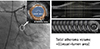
Patients underwent an IVUS examination before and after the stenting procedure. Patients received intracoronary nitroglycerin (200 µg) and the IVUS examination was performed using a commercially available system (CVIS; Boston Scientific Corporation, San Jose, CA, USA). The system consisted of a single-element 40-MHz transducer mounted on the tip of a flexible shaft rotating at 1800 rpm within a 2.6-F rapid exchange/common distal lumen imaging sheath. The examination was carried out with an automated pullback device at a rate of 0.5 mm/s. The images were digitized to perform morphometric analysis with commercially available planimetry software (echoPlaque, IndecMedical Systems, Santa Clara, CA, USA). We estimated the change in atheroma volume in a target lesion located more than 10 mm away from the stent and in the neointimal proliferation of the stent area. In one subset of patients, the lumen and vessel cross-section areas (CSAs) were measured at 1.0-mm increments from a point at least 10 mm away from both stent edges. Similarly, lumen and stent CSAs were measured throughout the stented segment at 1.0-mm increments. The atheroma CSA was calculated as the vessel CSA minus the lumen CSA (Fig. 1). The neointimal CSA was then calculated as the difference between the stent CSA and lumen CSA. The atheroma volume and neointimal volume were calculated by the Simpson's method. The atheroma volume index and neointimal volume index were calculated as the volume divided by the length of the measured atheroma, whereas percent atheroma volume was calculated as the ratio of atheroma volume to vessel volume×100.
Study endpoints
The primary endpoints included changes in atheroma volume index and percent atheroma volume from baseline in the target lesion, while secondary endpoints included changes in neointimal volume index and serum lipid profiles at the 12-month follow-up.
Statistical analysis
A sample size of 36 patients per group was calculated for an 80% power to detect a difference in the mean percent atheroma volume index of 4, assuming a standard deviation of 8 in the primary outcome variables and an alpha error of 5%, and a dropout rate of 10%. The D'Agostino Pearson test was used to evaluate for variable normality. Baseline descriptive data were expressed as mean±standard deviation for normally distributed continuous variables, as medians (interquartile ranges) for other continuous variables, and as percentages for categorical variables. The distributions of continuous variables were compared using the Student t-test or Mann-Whitney U test, as appropriate. Chi-square tests were used to compare the distributions of categorical variables. A p<0.05 was considered statistically significant. All statistical analyses were performed using the SPSS version 18.0 software package (SPSS Inc., Chicago, IL, USA).
Results
Baseline clinical and angiographic characteristics
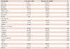
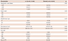
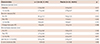
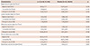
No differences were observed in the baseline clinical characteristics between the two groups. The mean ages were 59.6 and 60.7 years in the n-3 and placebo groups respectively, and more than 60% of patients were men. While the rate of stable angina was high in both groups, the incidence of non-ST-segment elevation MI was higher in the n-3 group; however, this difference was not statistically significant. No difference in the distribution of medications, including lipid-lowering, antiplatelet, or antihypertensive agents, between the groups (Table 1) was observed. Similarly, between the groups, there was no significant difference in the baseline angiographic characteristics (Table 2) and the type of implanted stents. The left anterior descending artery appeared to be the most commonly obstructed vessel.
Quantitative coronary angiographic analyses at baseline and at the 12-month follow-up
Quantitative coronary angiographic (QCA) data are presented in Table 3. Diameter stenosis at baseline was more significant in the n-3 group than in the placebo group (81.3% vs. 74.8%); however, all other post-procedural parameters were similar. No differences in late loss (0.54 mm, n-3 group; 0.46 mm, placebo group; p=0.438), or rate of in-stent restenosis (14.3%, n-3 group; 13.6%, placebo group; p>0.999), were observed between the two groups.
Intravascular ultrasound analyses at baseline and at the 12-month follow-up
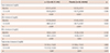
The results of the IVUS analysis at the 12-month follow-up are shown in Table 4. No significant decrease in atheroma volume was observed in the n-3 group compared with the placebo group, measured in terms of percent change in atheroma volume index (−12.65% vs. −8.51%, p=0.768) and change in percent atheroma volume (−4.36% vs. −9.98%, p=0.526). Similarly, neointimal volume (neointimal volume index; 7.84 mm3/mm, n-3 group vs. 4.94 mm3/mm, placebo group, p=0.154) did not differ between the two groups.
Changes in lipid profiles from baseline
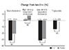
As shown in Table 5, lipid profiles at baseline were similar between the two groups. At the 12-month follow-up, the total cholesterol and LDL cholesterol levels significantly decreased in both groups (p<0.001). In addition, although HDL cholesterol levels increased significantly compared with the respective baseline values in the n-3 group (p<0.001), no significant differences in these levels were observed between the two groups. Triglyceride levels did not show a significant reduction between the two groups (Fig. 2) at the 12-month follow-up.
Discussion
The main results of this study can be summarized as follows: addition of ω-3 PUFA to statin therapy in CAD patients requiring stent implantation did not have any effect on atheroma volume index, change in percent atheroma volume, neointimal volume index, late loss, and in-stent restenosis rates. These findings suggest that the additive benefits of ω-3 PUFA on atherosclerosis regression are questionable.
Atherosclerosis, a major cause of CAD, is characterized by a series of processes which include endothelial dysfunction, lipoprotein accumulation, leukocyte recruitment by receptors or adhesion molecules such as vascular cell adhesion molecule (VCAM) intercellular adhesion molecule (ICAM), or p-selectin, and macrophage differentiation to lipid-laden foam cells that then form the atheroma. Furthermore, smooth muscle cell proliferation and migration, induced by pro-inflammatory cytokines such as tumor necrosis factor (TNF)-α, interleukin (IL)-1, or interferon (IFN)-γ, complicate atheroma evolution and increase the risk of CAD.9) Several studies1)2)3)4)5)10)11)12)13) have attempted to clarify the mechanisms of the atheroprotective effects of ω-3 PUFA. Apart from lowering the serum triglyceride level (a marker for several atherogenic lipoproteins or proinflammatory proteins promoting atherogenesis),9)10)11) ω-3 PUFA improves the endothelial function and displays antithrombogenic effects.1)2)11) Moreover, it reduces the levels of adhesion molecules and inflammatory mediators by the regulation of gene expression or transformation to downstream metabolites.12) It is believed that through these mechanisms, ω-3 PUFA increases the atherosclerotic plaque stability.2)3)13) This view is supported by results from a previous study using IVUS analysis by Amano et al.,4) in which a low serum ω-3 PUFA level was associated with the development of vulnerable plaques, suggesting a higher risk of acute coronary syndrome.4) Other study5) demonstrated that low serum ω-3 PUFA was associated with atheroma progression in patients who achieved very low levels of LDL cholesterol during statin therapy.5) In contrast, in this study we hypothesized that elevation of serum ω-3 PUFA by ω-3 PUFA supplementation could induce the regression of coronary atherosclerosis. Nakajima et al.14) reported that orally administrated EPA induces the regression of atherosclerosis in LDL receptor-deficient mice, and that this effect was possibly mediated by alteration of dendritic cell functions and decrease in circulating T-lymphocytes. In our study, CAD patients received 3 g of ω-3 PUFA in addition to statin therapy; however, we did not observe regression of coronary atherosclerosis or prevention of neointimal proliferation as assessed by IVUS. Although this finding suggests that supplementation with 3g of ω-3 PUFA is not associated with regression of coronary atherosclerosis, ω-3 PUFA serum levels prior to ω-3 PUFA supplementation, were not measured in this study. As no data on the optimal level of serum ω-3 PUFA necessary for regression of coronary atherosclerosis exists, we must consider the possibility that the 3 g ω-3 PUFA dose chosen was not adequate.
The cardiovascular protective roles of ω-3 PUFA can also be explained by mechanisms other than the above mentioned atheroprotective effects, including the suppression of cardiac arrhythmias, reduction of myocardial oxygen consumption, improvement of cardiac filling and myocardial efficiency, regulation of insulin resistance, and decrease in heart rate or blood pressure.15)16)17) Based on this information, results from meta-analyses have demonstrated a significant decrease or trend toward a decrease in mortality from CAD in patients with or without underlying cardiovascular disease.3)18)19)
However, in contrast to earlier studies, no significant positive additive effects of ω-3 PUFA on atheroma regression were observed by IVUS analysis in the present study. Possible explanations for this discrepancy are as follows: first, medication usage, especially statins, differed between studies. As statins are known to significantly decrease the relative risk of morbidity and mortality from cardiovascular diseases,20) they are considered the first-line therapy for primary or secondary prevention. However, although results from the GISSI-Prevenzione trial21) conducted in the mid-1990s showed benefits from statin treatment, only 5% of the eligible patients received statin therapy, whereas no benefit was seen in two recently conducted landmark trials, i.e. the ORIGIN and the OMEGA trials,8)9) in which more than 50% and 80% of the patients received statin therapy, respectively. Moreover, while the effects of ω-3 PUFA on lipid profiles are mainly associated with triglycerides, a previous study showed that atheroma regression was related to LDL cholesterol levels.22) Considering the effects of statins, we hypothesized that adding ω-3 PUFA to statin therapy could have additive benefits. However, IVUS analysis showed that the use of ω-3 PUFA, compared to placebo, did not affect the regression of atherosclerosis, and only caused a slight decrease in triglyceride levels. Second, dietary differences in the consumption of fish containing abundant ω-3 PUFA may have an influence on serum ω-3 PUFA levels. For instance, the omega-3 index is relatively high in Korea because of a fish-rich diet.23) Furthermore, as the validity of a dose-response model for studying the cardiovascular effects of ω-3 PUFA remains controversial even in meta-analyses, the cut-off dose is currently undefined.19)24) In the present study, we hypothesized that a high dose (3 g/day) of ω-3 PUFA could have additive effects, but the possibility that patients in the placebo group had higher ω-3 PUFA baseline levels because of higher fish consumption, may have prevented induction of additive effects by the combined treatment. Third, the study populations differed between the studies. Previous trials, in which significant cardiovascular benefits of ω-3 PUFA were reported, included patients with a high risk of cardiovascular disease. Moreover, the GISSI-Prevenzione trial21) enrolled patients with MI within the previous 3 months, the GISSI-Heart Failure trial25) included patients with chronic congestive heart failure, and the ESPRIT trial26) included patients with traditional cardiovascular risk factors and hypertriglyceridemia>200 mg/dL. However, in this study, we excluded patients with acute MI, left main coronary artery disease, severe heart failure with LVEF<40%, uncontrolled hypertension, or diabetes. Therefore, our patients had a relatively low risk of future cardiovascular events, and the serum triglyceride level was an average of 150 mg/dL at baseline. Despite results from previous studies,27)28) the cardiovascular benefits of ω-3 PUFA in patients with relatively low risk of cardiovascular disease appears difficult to prove. Furthermore, one previous study reported that therapy with ω-3 PUFA did not reduce in-stent restenosis.29) In our study, ω-3 PUFA had no effect on late loss, in-stent restenosis, and neointimal volume index. Therefore, we consider that the anti-inflammatory effects of ω-3 PUFA are not strong enough to reduce neointimal proliferation.
Limitations
First, although we designed this study to investigate the benefits of adding ω-3 PUFA to statin therapy, we did not compare the differences between the statin group and the placebo group in the same patient population. Moreover, statins prescribed in this study were limited to only atorvastatin and rosuvastatin, since in actual clinical settings, these are the most commonly used statins. Second, we did not measure ω-3 PUFA level in serum, red blood cells, or some other long-term marker of its enrichment prior to supplementation and did not control for dietary fish consumption. Therefore, the optimal levels of ω-3 PUFA needed to induce regression of coronary atherosclerosis are unknown. Third, as the serum triglyceride level at baseline was relatively low (average of 150 mg/dL), no significant reduction in serum triglyceride level was observed after 3g of ω-3 PUFA supplementation. Therefore, the additive effect of ω-3 PUFA was not definite in our study. Fourth, this study was single-blinded. However, this study design did not seem to affect the outcomes since all the patients in both groups completed a follow-up period, and a follow-up study was performed at the same time.
Despite these limitations, this study provides unique information, in that we used IVUS to examine the potential additive effects of ω-3 PUFA on atherosclerotic plaques in CAD patients receiving statin therapy.
 XML Download