

 PDF
PDF ePub
ePub Citation
 Print
Print


Introduction
Azygos vein aneurysm presenting as a mediastinal mass is a rare disease entity. No more than 50 cases have been reported to date, with age ranging from 19 years to more than 70 years. There have been no previous reports of azygos vein aneurysm being diagnosed before 18 years of age. Although most cases are found asymptomatic at diagnosis, few case reports introduce complicated azygos vein aneurysms in need of surgical intervention. No ruptured azygos vein aneurysm has so far been reported, except for a case of a pseudoaneurysm caused by trauma. We report a case of azygos vein aneurysm in infancy, complicated with pulmonary thromboembolism, who underwent surgery to remove the aneurysm. To date, no report could be found about an azygos vein aneurysm diagnosed in infancy, and the number of cases of complicated azygos vein aneurysms requiring surgical intervention is limited throughout literature, for all ages.
Case
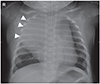
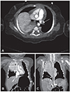
A 3-month old boy was transferred to our hospital due to the sudden onset of tachycardia and cyanosis. The baby had previously been diagnosed as a lymphangioma of posterior mediastinum on prenatal fetal ultrasound study, and was routinely followed up in another hospital, without any medication. The day before admission to our hospital, the parents took the baby to an emergency center because of a sudden onset of cyanosis and dyspnea. At the time of arrival, his heart rate was 200/min, and the peripheral capillary oxygen saturation (SpO2) was 80%, with no response to bag-valve mask ventilation. His lung sounded clear without adventitious sounds. His heart rate was 188/min, but no cardiac murmur was heard. His chest x-ray showed a soft tissue-density mass at the right hemithorax and paraspinal area (Fig. 1). He soon showed a seizure-like movement, which was not controlled by midazolam, lorazepam, or phenobarbital. A brain non-contrast computed tomography (CT) did not show any abnormal findings, and a contrast chest CT scan revealed a cystic lesion (about 10 cm in size) on the right thorax, with a thromboembolism in the left proximal pulmonary artery (Fig. 2). He was immediately intubated and referred to our hospital while on heparinization, for further evaluation and management.
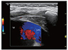
He was directly admitted to the Pediatric Intensive Care Unit with stable vital signs. His body gauge was 66 cm (75-90 percentile) and 9.6 kg (>97 percentile). His initial heart rate was 188/min, blood pressure was 84/49 mmHg, respiration rate was 56/min, and SpO2 was 100% with ventilator support (fraction of inspired oxygen: 0.4). His lungs sounded clear without any adventitious sounds, and his heart sounded normal. His abdomen was soft and not distended, but a mild hepatosplenomegaly of 1 finger breadth was detected. Echocardiography, chest and abdomen ultrasound were done and reviewed along with the initial chest CT (Figs. 2 and 3). Thrombus was visible in the left pulmonary artery; a connection was found between the previously presumed lymphangioma-like mass lesion and the superior vena cava (SVC), and the mass lesion and azygos vein, thus supporting the diagnosis of azygos vein aneurysm rather than lymphangioma. Within the next 12 hours, the SpO2 level went down to 20%, and emergency thromboembolectomy and disconnection of the mass and vessels were performed. Multiple thrombi were removed from the left pulmonary artery using a thoracoscope, and two connections (between the mass and SVC) were found and ligated. A lower smaller connection was located directly below the proximal innominate vein, while an upper larger connection was found at the usual drainage level of the azygos vein.
Post-operative vital signs were stable, but extubation failed twice within the next 20 days due to heavy secretions, chest retraction, and carbon dioxide accumulation after every extubation trial. Bronchoscopy showed a narrowing of the tracheal airway, reflecting the remaining mass effect of the aneurysm. A second operation was done within 24 hours, after an airway evaluation by bronchoscopic examination, to remove the remaining mass. Resection of the mass was successfully performed after reducing the total volume of the mass, by ligating the multiple arterial and venous connections to the mass, including the connection between the mass and the azygos vein. The expansion of the right lung after surgery seemed normal. Five days later, the boy was extubated without complications, and he was discharged 2 weeks later. The follow up chest CT scan taken before discharge showed complete removal of the vascular mass in the posterior mediastinum, and non-visualization of the right upper lobar bronchus with no evidence of pulmonary thromboembolism.
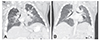
No cardiopulmonary symptoms developed in the follow-up outpatient clinic visits after discharge. The chest contrast CT 6 months later showed complete removal of the vascular mass with demonstrable right upper bronchus, which was previously suspected to be collapsed due to the aneurysm (Fig. 4). Echocardiography showed no evidence of dilated vessels or pulmonary hypertension, and a 24 hour holter test was normal.
Discussion
To our knowledge, this is the first pediatric case report of a complicated azygos vein aneurysm requiring surgical intervention. The youngest case reported before this was an asymptomatic 19 year old male, whose diagnosis was confirmed through surgical intervention in 1961.1) The presenting case showed that an azygos vein aneurysm and its complications can occur very early in life, or potentially in all age groups, alerting physicians and pediatricians to suspect the diagnosis in mediastinal mass lesions.
Early diagnosis is quite important, as seen in the above case. If echocardiography or abdomen ultrasound was performed before the development of symptoms, the diagnosis might have been confirmed earlier, and early intervention might have been possible before the progression to pulmonary embolism. Contrast enhanced dynamic CT2) and magnetic resonance imaging3) are helpful non-invasive methods to confirm the diagnosis. However, chest ultrasound and echocardiography can provide additional information in revealing the communication of the aneurysm to the superior vena cava, as shown in the above case. Invasive modalities, including venography or biopsy, are not usually considered once the diagnosis is made.4)
Surgical intervention was inevitable in the presenting case for the relief of pulmonary thromboembolism. However, the necessity of the treatment for an asymptomatic azygos vein aneurysm at the time of diagnosis remains controversial. In some reports, the conservative management with periodic imaging follow-up showed no adverse outcomes.2) However, based on the case reports of complications, other studies stress the need for treatment in all azygos vein aneurysms to prevent future complications such as thrombosis leading to pulmonary embolism, mass effect, or venous rupture,2)3)4)5)6)7)8) including the possibility of sudden thrombosis development.9) Ko et al.6) have proposed the possibility of selective treatment, suggesting that saccular aneurysms are less stable and more complicated than fusiform aneurysms.
Although surgical intervention remains the treatment of choice for most azygos vein aneurysms,5)8) other modalities are also promising. Video assisted thoracoscopic resection is an attractive method of surgery in selected cases with no thrombus,10) and thrombolytic therapy, stent-graft replacement, and transcatheter occlusion have also been tried and proved effective in limited cases.11)12)13) Further studies are necessary for comparison of new modalities to surgical intervention in specific situations.
There are some debatable points in the presenting case regarding the management of a complicated azygos vein aneurysm. While further studies might have revealed the azygos vein aneurysm before the thromboembolism development, it remains unclear whether surgical intervention was necessary at the time of diagnosis. Although the huge size of the mass lesion, which reflects a higher possibility of impending complications, favors the surgical intervention, the low incidence of complications and the risk of surgical intervention supports the decision of close observation. Close follow up with appropriate imaging modalities would be a reasonable choice for management, with the guardians being given sufficient warnings and cautions about the possible complications.
In conclusion, this case is the youngest case of a complicated azygos vein aneurysm presenting a life-threatening pulmonary thromboembolism in infancy. Differential diagnosis for mediastinal mass in infancy consists of various disease entities. However, vascular lesion can be one possibility, and a contrast enhanced CT scan or echocardiography can help in confirming the diagnosis. The intervention, usually surgical, is necessary if complications exist, but management remains controversial if there are no complications. Further studies and individualization of the situations are necessary for appropriate management.
 XML Download