

 PDF
PDF ePub
ePub Citation
 Print
Print


Introduction
Symptoms of cardiac involvement of tumor result from the location and impingement on adjacent structures. Conduction disturbances due to several kinds of cardiac tumor such as primary rhabdomyosarcoma,1) malignant melanoma with cardiac metastasis,2) cardiac hemangioma3) and metastatic adenocarcinoma of lung4) were reported previously. Mesotheliomas of the atrio-ventricular (AV) node also cause heart block and sudden death.5) However, cardiac involvement of leiomyosarcoma is very rare. We reported a case of cardiac metastasis of leiomyosarcoma complicated with bradyarrhythmia and tachyarrhythmia.
Case
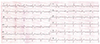
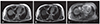
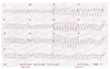
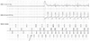
A 54-year-old man was referred to our electrophysiology laboratory because of dizziness and dyspnea. He was diagnosed with leiomyosarcoma of the right lower leg and received wide excision at another hospital 5 years ago. Later, he underwent several operations due to recurrent pulmonary lesions. Histological examination confirmed metastasis of leiomyosarcoma. Recently, the patient was in good physical condition for 2 years and no abnormal findings were detected on the surface electrocardiography (ECG) 2 years prior. However, the current ECG revealed complete AV block and idioventricular escaped rhythm of bifascicular block morphology suggesting infrahisian block (Fig. 1). Chest computed tomography revealed multiple pulmonary lesions and significant thickening of interventricular septum. The patient underwent cardiac magnetic resonance imaging, which revealed huge interventricular mass (67×35 mm) from base to apex with low signal intensity, similar to the myocardium on the T1-weighted image (Fig. 2A) and mild higher signal intensity, as compared with the myocardium on the T2-weighted image (Fig. 2B). After enhancement with gadolinium, the tumor showed peripheral enhancement (Fig. 2C). Trans-thoracic echocardiography revealed normal left ventricular ejection fraction (LVEF 60%) and no hemodynamic compromise. Dual chamber permanent pacemaker was implanted on the second day of admission and we placed a right ventricular (RV) lead toward free wall of RV apex that was confirmed by fluoroscopy and echocardiography because of tumor invasion in the septum of RV apex. There were no procedure-related complications, however, ventricular tachycardia (VT) developed 3 days later. Twelve-lead ECG showed wide QRS complex tachycardia, left bundle branch block pattern with left superior axis morphology and late transition, which is compatible with VT from RV apex (Fig. 3). Administered amiodarone and ß-blocker suppressed further events of VT and the patient was discharged uneventfully. He received pazopanib for palliative chemotherapy, which was stopped due to hepatic toxicity and poor performance status. The patient passed away after 3 months of pacemaker implantation due to progression of underlying disease and multiple organ failure.
Discussion
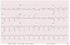
There are only a few sporadic case reports of arrhythmic presentation in patients with cardiac involvement of leiomyosarcoma. Atrial fibrillation due to metastasis to pulmonary vein and left atrium,6) ectopic atrial tachycardia with right atrial leiomyosarcoma7) and VT due to local tumor growth in the right ventricular outflow tract8) were reported previously. This was the first case report of a large metastatic mass of leiomyosarcoma located on the entire interventricular septum causing complete AV block. Furthermore, there are no previous reports of cardiac involvement of tumor that caused both bradyarrhythmia and tachyarrhythmia. The patient was treated with implantation of permanent pacemaker for the management of complete AV block and anti-arrhythmic drug suppressed VT successfully. Pacemaker mediated tachycardia or pacemaker-induced VT was considered because of subsequent development of VT after pacemaker implantation without prior history of VT. However, 12-lead QRS morphology of VT was different from that of paced ventricular rhythm (Fig. 4) and apical septum of RV due to tumor invasion was suspected as the origin. Pacemaker interrogation excluded pacing-induced VT or pacemaker-mediated tachycardia (Fig. 5). We concluded that VT was another arrhythmic manifestation of cardiac involvement of leiomyosarcoma.
There was no histopathological diagnosis of cardiac lesion, however, we diagnosed the mass as a malignant metastasis because of the simultaneous similar pattern of widespread metastatic lung lesions with prior histopathologic confirmation of metastatic malignant leiomyosarcoma. Soft tissue sarcomas are resected surgically whenever feasible. In our patient, complete surgical resection of cardiac mass was not feasible because of infiltrative growth in the entire interventricular septum. Pacemaker implantation and anti-arrhythmic medications were started for symptomatic relief. Upgrade to implantable cardioverter defibrillator (ICD) was considered regarding underlying structural substrate for VT. However, VT was well controlled with anti-arrhythmic drug and overall mean survival time in patients with metastasizing soft tissue sarcoma is approximately only 10 months despite therapy.9) Thus, upgrade to ICD was not performed and our patient passed away 3 month later due to disease progression
Cardiac metastasis should be considered when the patient with previously known leiomyosarcoma complains of cardiac symptom. Clinicians should be aware that both bradyarrhythmia and tachyarrhythmia could develop in patients with cardiac metastasis of malignant leiomyosarcoma.
 XML Download