

 PDF
PDF ePub
ePub Citation
 Print
Print


Introduction
Cardiac catheterization was used for diagnosing structural heart disease (SHD) before the development of modern echocardiography. Despite the development of cardiac computed tomography and magnetic resonance imaging since the early 2000s that has allowed safer and more accurate diagnosis of SHD, cardiac catheterization continues to play an important role in assessing the hemodynamic status of SHD. Recently, because of more advanced therapeutic interventions for SHD and the increased incidence of adult congenital heart disease, the number of cardiac catheterization procedures and associated transcatheter treatments for SHD has increased.1) Cardiac catheterization technique and medical environment varies considerably between catheterization laboratory centers and countries; this could cause differences in complications of cardiac catheterization. Centers in western countries have reported various complications of cardiac catheterization since the mid-1970s with the overall complication rates ranging from 8.8–24%.2)3)4)5)6)7)8)9)10) However, there is no data on overall complications of cardiac catheterization for SHD in Korea and the associated risk factors. The aim of this study was to determine the category, frequency, and associated risk factors of overall and severe complications of cardiac catheterizations for SHD performed over a recent 10-year period.
Subjects and Methods
Study population
A retrospective analysis was performed using the data collected from patients who underwent cardiac catheterization at the Seoul National University Children's Hospital from January 2004 to December 2013. Cases in which electrophysiological study (EPS) procedures were performed primarily to evaluate arrhythmias or with radiofrequency catheter ablation were excluded from the study. However, cases of EPS concurrent with cardiac catheterization for SHD were included in the study.
The Institutional Review Board of Seoul National University Hospital approved the study, and informed consent was waived because of its retrospective nature.
Data collection
All electronic and paper chart records were assessed to obtain precise information, including age at procedure, procedure date, gender, weight, admission ward (intensive care unit [ICU] or general ward), underlying SHD, usage of antithrombotic agent, prothrombin time (PT)/activated partial thromboplastin time (aPTT) before procedure, total number of cardiac catheterizations in each patient, complications within 24 hours of the procedure, sedation method, procedure duration, total fluoroscopic time, amount of contrast dye used during the procedure, and name of the intervention performed during the procedure.
Grouping and definition
Age at procedure was grouped as <1 month, 1–12 months, 1–8 years, 8–15 years, 15–20 years, and ≥20 years. SHDs were graded as mild, moderate, and severe, according to the Task Force 1 of the 32nd Bethesda Conference of the American College of Cardiology in 2001.11)
Despite usual cessation of drugs 1 week before the catheterization date, only use of aspirin or clopidogrel prior to the procedure was defined as anti-platelet agent use and any use of warfarin or heparin prior to the procedure was defined as anti-coagulation agent use.
Interventional catheterization consisted of procedures involving manipulative therapy (myocardial biopsy, ballooning, coil embolization, device closure, stent insertion and dilatation, etc.). Other diagnostic studies were performed to evaluate anatomic structure and/or assess hemodynamic status of each heart disease.
In this study, general anesthesia was defined as deep sedation with respiratory support using a mechanical ventilator; and intravenous (IV) anesthesia was defined as mild to moderate sedation with IV drugs such as ketamine or midazolam without an artificial airway.
Procedure time was the duration between the start time, defined as arterial or venous catheter insertion, and the finish time, defined as the removal of the catheter or departure from the procedure room without catheter removal.
Complications within 24 hours of procedure were classified into 16 categories, and each complication was grouped as mild, moderate, or severe complication (Table 1). Severe complications included death, cardiac arrest accompanied by cardioversion, pacemaker insertion, chest compression, cerebrovascular embolization, anaphylaxis, emergent intubation, or events that required any surgery. Minor complications were transient events, and mostly resolved without specific treatment.
Statistical analyses
Descriptive data were presented as means with standard deviation or medians with ranges, whereas categorical variables were presented as proportions. Each procedure was identified and analyzed; however, the total number of catheterizations was calculated by cumulative procedure number in each patient from 2004. If there were multiple interventions or complications in a procedure, the number of procedures applied to the intervention types and complication types overlapped.
All complications were tabulated according to the complication categories and severity, and the rate of complication was reported as a percentage of the total procedures. The relationships between diverse variables and overall and severe complication risks, separately, were analyzed using univariate logistic regression analysis.
Data manipulation and statistical analyses were performed with SPSS 21.0 for Windows (SPSS Inc., Somers, NY, USA) and Excel 2010 (Microsoft, Santa Rosa, California, USA). Observations with a p<0.05 were considered statistically significant.
Results
Baseline characteristics
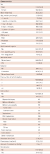
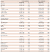
Demographic data and clinical characteristics of patients at the time of procedures were described in Table 2. A total of 2071 cardiac catheterizations, performed in 1609 patients (some patients had undergone several cardiac catheterization procedures), were identified during the 10-year study period. The median age was 5.5 years (range, 1 day–59 years), and the mean body weight was 26.2±20.5 kg. According to the underlying SHD, 735 cases were classified as severe, 1115 as moderate, and 221 as mild. There were 1279 (61.8%) diagnostic catheterizations, 237 balloon dilatations for peripheral vessels, 204 device implantations, 156 coil embolizations, 99 balloon valvuloplasties, 63 myocardial biopsies, 23 stent insertions, 22 septostomies, 6 stent dilations by balloon, and 1 urokinase infusion into the coronary artery. Among cases of multiple interventions in a single procedure, there were 10 cases of balloon dilation and stent insertion, 6 of balloon dilatation and coil implantation, 2 of coil implantation and patent ductus arteriosus (PDA) device implantation, 1 of pulmonary valvuloplasty and coil implantation, and 1 of stent insertion and septostomy.
During the 10-year study period, 1303 patients underwent the procedure once, 230 patients twice, 44 patients thrice, 16 patients 4 times, and 16 patients≥5 times. The group in which the procedure was performed >5 times underwent either endomyocardial biopsy after heart transplantation or ballooning in pulmonary stenosis. There were 172 (8.3%) cases admitted to the ICU, and 942 (54.5%) patients were males. For sedation, 399 (19.9%) cases were performed under local analgesic anesthesia, 1474 (71.1%) cases were managed with IV sedation, and 198 (9.6%) cases were performed under general anesthesia. Anti-platelet agents were administered in 15.1% of procedures, and anti-coagulation agents in 5.2% of procedures. Mean PT level was 1.13±0.25 international normalized ratio (INR), and mean aPTT was 39.62±8.24 seconds. In the anti-platelet treated group, mean PT INR was 1.13±0.17 and mean aPTT was 39.5±7.2 seconds. In the anti-coagulant treated group, mean PT INR was 1.65±0.63 and mean aPTT was 46.0±17.3 seconds. On the other hand, in the treated group without anti-thrombotic agent, mean PT was 1.10±0.16 and aPTT was 39.2±7.3 seconds. Mean procedure time was 57.4±31.6 minutes, mean fluoroscopic time was 17.2±12.5 minutes, and mean amount of contrast dye per weight was 2.5±1.7 cc/kg.
Complications
A total of 314 patients had a total of 335 complications that were grouped into 16 categories and 3 severity scales (Table 1). The number of complications (Table 3) was overlapped in case of multiple complications. The percentage was calculated by dividing by the total procedure number, 2071.
Among the overall complications, 264 cases were classified as mild, 47 cases as moderate, and 24 cases as severe. The calculated incidence of overall complication was 16.2%, and severe complication was 1.15%. For the diagnostic catheterization alone, overall and severe complication rates were 14.4% and 1.0%; and for the therapeutic catheterization, overall and severe complication rates were 16.4% and 1.4%.
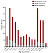
The most frequent overall complication was fever, followed by gastrointestinal complications such as vomiting, respiratory complication, arrhythmia, vascular thrombus, and vascular bleeding. The most common mild complications were fever (n=94), nausea and/or vomiting (n=51), and desaturation requiring mask oxygen supply (n=38), the same order as the overall complication. The most common severe complications were arrhythmias requiring cardioversion or pacemaker (n=10). There were 4 cardiac catheterization-related deaths and 5 chest compression events. There were 4 patients who required surgical intervention due to the severe complications i.e., vascular thrombus in the pulmonary artery after Glenn shunt, cardiac perforation during myocardial biopsy, end hole catheter fracture, and atrial septal defect device embolization into the aortic arch. There was 1 case of emergent intubation during catheterization due to respiratory instability, and 1 case of right middle cerebral artery infarction that required thrombectomy. Severe complications mainly occurred in patients <3 years of age or in patients >10 years of age, but were most frequent in infants (Fig. 1).
Risk factors for complications of cardiac catheterization
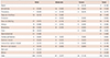
Risk factor analysis for complications was described in Table 4. The variables that showed significant differences in the occurrence of both overall and severe complication were anticoagulant use (odds ratio [OR] 1.83, p=0.012 and OR 6.45, p<0.001, respectively), PT (OR 2.30, p<0.001 and OR 5.99, p<0.001, respectively), general anesthesia use (OR 1.84, p=0.014 and OR 5.31, p=0.015, respectively), and procedure time (OR 1.01, p<0.001 and OR 1.02, p<0.001, respectively). When anticoagulants such as heparin or warfarin were used prior to the procedure, the number of overall and severe complications was 1.83 and 6.45 times higher, respectively, than when antithrombotic agents were not used (p=0.012 vs. p<0.001). However, use of antiplatelet agents such as aspirin or clopidogrel resulted in no significant difference in the complication rate. Compared with procedures performed without sedation, the total number of complications significantly increased by 1.58 times (p=0.011) in procedures involving IV sedation and by 1.84 times (p=0.014) in procedures performed under general sedation. The rate of severe complication increased by 1.12 times (p=0.859) and 5.31 (p=0.015) times, respectively.
Patients with severe underlying SHD had a significantly higher risk of overall complication than those who had mild to moderate SHD (p=0.012). As the number of catheterization procedures increased in each patient, the patient had a higher risk of overall complications (p=0.035). The probability of overall complications in catheterization >3 times was 1.70 times higher than the first or second catheterization (p=0.009).
Although there was no significant difference in the overall rate of complications depending on age groups, infants <1 month appeared to have a 3 times higher risk of severe complication than adults >20 years old (OR 3.00, p=0.089). The severe complication rate was significantly high in the procedures with high serum aPTT, ICU admission, and concomitant EPS procedure.
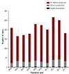
There was no significant difference in the incidence of complication incidence according to gender, year of procedure (Fig. 2), presence of pulmonary hypertension, type of procedure, diameter of arterial or venous catheter, and amount of contrast dye injected during the procedure.
Mortality cases
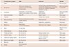
There were 4 deaths during the 10 years of study, indicating a mortality rate of 0.19%. The details of mortality cases were described in Table 5. Three patients died after the first catheterization at <2 months of age, of circulatory arrest during the peri-procedural period.
Two mortality cases were neonates with hypoplastic left heart syndrome that needed emergent PDA stenting. One was a 28 day-old infant who had bilateral pulmonary artery banding while awaiting the Norwood operation, due to necrotizing enterocolitis. Although the septic condition improved, hypotenstion, bradycardia, and oliguria occurred because of PDA constriction. Therefore, we performed emergent PDA stenting. However, hypotension, bradycardia, and oliguria persisted even after PDA stenting, which led to death at 8 hours after the procedure. The other hypoplastic left heart syndrome also had bilateral pulmonary artery banding and ileostomy due to necrotizing enterocolitis. We planned emergent PDA stenting on aggravated tachypnea and desaturation possibly due to the pulmonary edema. During the cardiac catheterization, cardiopulmonary resuscitation (CPR) was required thrice for bradycardia and desaturation. The patient died at 3 hours after femoral vein puncture.
The other neonate had critical aortic valve stenosis, which was the indication for emergent balloon aortic valvuloplasty at 9 days of age. Severe hypotension occurred during the advancement of catheter into the ascending aorta through the right carotid artery. CPR was followed and he expired 2 hours after femoral vein puncture. There was neither extravascular bleeding nor cardiac tamponade.
The last mortality case was an infant with pulmonary atresia with intact ventricular septum when diagnostic catheterization including right ventricular angiography was performed before the Glenn shunt surgery. The patient showed sudden bradycardia 4 hours after catheterization (presumably due to thromboembolic event) that needed CPR, followed by extracorporeal membrane oxygenation insertion. After 26 hours from cardiac catheterization, the patient died of persistent metabolic acidosis and ventricular tachycardia.
Discussion
In this study, we analyzed all cardiac catheterization procedures performed for SHD in a single center during the latest decade, in order to identify the type and frequency of complications and determine the associated risk factors.
Previous studies have reported overall complication rates ranging from 8.8 to 24%, and mortality rates ranging from 0.14 to 2.7%.2)3)4)5)6)7)8)9)10) The overall complication rate observed in this study was 16.2% including minor complications such as fever, nausea, and vomiting, which accounted for 43.3% of all complications in our study, but were excluded in the previous studies. In our study, the incidence of severe complications was only 1.15% and the mortality rate was only 0.19%. The complication rates were higher in the therapeutic intervention group than the diagnostic catheterization group.
To date, few studies have reported complications in pediatric cardiac catheterization. An institute in Toronto, Canada reported complications in pediatric cardiac catheterization procedures performed between 1987 and 1993 and between 1994 and 2006; major complications occurred at a rate of 2.1% and 1.8%, respectively, and mortality rate was reportedly 0.14% and 0.25%, respectively.2)12) Japanese pediatric interventional cardiology data from 2004–2008, including 8446 therapeutic interventional procedures, but excluding diagnostic procedures, indicated that major complications occurred at a rate of 3.7% and mortality at a rate of 0.13%.9)
Collectively, the data from the Toronto study, the Japanese multi-center study, and our study showed that major or severe complications in pediatric cardiac catheterization occurs at a rate of 1–2% and mortality at a rate of 0.13–0.25%; occurrence of severe complications and mortality is likely to vary according to the skill of the doctor, the equipment of the center, and the proportion of therapeutic catheterization performed.
The prevalence of complications in each category
The complication rate in each category varies across reported studies. While vascular thrombosis was the most common mild and overall complication in previous studies,2)12)13)14)15) our study revealed that fever was the most common complication, followed by nausea, vomiting and respiratory difficulty. For this reason, we included the aforementioned complications in the overall complications associated with cardiac catheterization. Compared with the Toronto study wherein procedures were mainly performed under general anesthesia,2) our study showed more respiratory complications because of the use of general anesthesia in only 9.6% of procedures; most procedures (71.1%) in our study were completed by IV anesthesia that has a risk of desaturation needing oxygen support or positive pressure ventilator support during the procedure.16) Nevertheless, the respiratory complications reported in our study were mild to moderate except in 1 case.
As in the previous study,12) the most common severe complication was arrhythmia (0.48%), followed by chest compression and death. Other studies showed that younger patients were susceptible to complications during the peri-procedural period;2)3)4)5)6)7)8)10)12) likewise, all 4 deaths in our study occurred in patients aged ≤6 months.
Risk factors
Previous studies reported that the risk factors of complication in cardiac catheterization include younger age, larger sheath and catheter, urgent procedure, long procedure duration, prior heparin use, more contrast dye use, the presence of pulmonary hypertension, therapeutic procedures, and the procedure year.2)3)4)5)6)7)8)12)14)16)17)18)19)20)21)22)
In addition, previous studies reported that younger patients were more likely to experience complication;2)3)4)5)6)7) in agreement, our study showed that the incidence of severe complications was significantly greater (3-fold) in patients aged <1 month.
Using anticoagulants before the procedure based on the serum PT, was a significant risk factor for both overall and severe complication. Additional analysis of the bleeding complication showed association according to the antithrombotic agent categories i.e., antiplatelet (OR 1.92, p=0.266), and anticoagulant (OR 10.39, p<0.001).
Procedure time was an important risk factor for both overall and severe complications, in our study. As procedure duration exceeded 10 minutes, overall complications occurred 10% more frequently and severe complication occurred 20% more frequently; the difference was significant.
In our study, procedures performed under general anesthesia were associated with more overall and severe complications than procedures performed under local analgesic anesthesia. Because we performed general anesthesia selectively in cases of severe SHD or in neonates mainly for therapeutic purpose, general anesthesia itself was not a predictable risk factor for complication in our study.
In cardiac catheterization with concomitant EPS, severe complications significantly increased, but there was no significant difference in overall complications. During the study period, we performed aggressive right ventricle pacing in patients with cyanotic SHD to induce ventricular tachycardia and 4 patients developed ventricular tachycardia or atrial flutter, which required direct current cardioversion.
Our study results suggested that low body weight, severe underlying SHD, increasing number of catheterizations, and longer procedure time increased mild to moderate complications. There was no significant difference in the annual prevalence of complications.
Limitations of this study
This retrospective study was performed using a complication database, which depended on the physician's commitment to completing the forms after each catheterization. Hence, despite our attempts to identify all complications from the medical records including fever, gastrointestinal complications that were not included in previous studies, minor data losses were possible.
The heterogeneity of SHD was another possible limitation since it varies according to medical center and country. We tried to categorize various characteristics such as SHD severity, patient age, admission ward, and number of catheterizations as the risk factors to overcome the limitation.
Conclusion
In conclusion, cardiac catheterization in SHD is increasing and becoming more complex currently, because of the increasing incidence of therapeutic catheterization in most advanced centers. This has led to persistent complications despite the preventive efforts. In our study, severe complications were associated with infancy, anticoagulation use before procedure, ICU admittance, longer procedure time, and concomitant EPS procedure. Careful patient selection for therapeutic catheterization and improved technique and management during the peri-procedural period are required to reduce complications in cardiac catheterization for SHD.

 XML Download