

 PDF
PDF ePub
ePub Citation
 Print
Print


Introduction
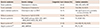
Congenital heart disease is the leading cause of birth defects, and is an important cause of morbidity and mortality during infancy and childhood. Congenital heart disease is a multifactorial disorder associated with both genetic and environmental influences. Approximately 30% of congenital heart disease is thought to be related to genetic syndromes accompanied by extra-cardiac anomalies.1) Recent advances in molecular genetic techniques have found evidence of the role of genetic factors in the development of congenital heart disease (Table 1). Understanding genetic etiology in a certain patient with both congenital heart disease and other anomalies can help clinicians to effectively plan a patient's surgical and medical management and their follow-up. Here, common genetic syndromes associated with various types of congenital heart disease are introduced with a brief review of their respective genetic backgrounds. The following are discussed: Down syndrome, Turner syndrome, 22q11 deletion syndrome, Williams syndrome, and Noonan syndrome.
Genetic Syndromes Diagnosed by Conventional Karyotyping
Down syndrome
Down syndrome is a chromosomal abnormality caused by the presence of a third copy of chromosome 21 and is manifested by characteristic facial dysmorphism, various congenital anomalies, including cardiac or gastrointestinal defects, variable degrees of intellectual disability, hypotonia, and joint laxity. The overall prevalence of Down syndrome has been estimated to be 1/700 live births,2) although it is highly dependent on maternal age and has decreased significantly as a result of routine prenatal testing.
Conventional karyotyping is indicated in all Down patients to confirm the diagnosis. Approximately 95% of patients are standard trisomy 21, 46, XX, +21 or 46, XY, +21. The remaining 5% of patients have Down syndrome resulting from Robertsonian translocations, which can be familial.
Congenital heart disease occurs in 40-50% of Down patients and is an important determinant of survival. Various cardiac malformations can be associated with Down syndrome, however atrioventricular canal defects, atrial and ventricular septal defects, and tetralogy of Fallot are commonly observed.3) As a result, echocardiograms should be performed in all patients at diagnosis, regardless of whether a fetal echocardiogram was performed.4) It is well known that pulmonary hypertension develops more often and earlier in patients with congenital heart disease and Down syndrome. In addition, the incidence of persistent pulmonary hypertension of a newborn is increased 10-fold in those with Down syndrome. Nevertheless, many epidemiological studies have reported that Down syndrome does not increase surgical mortality and morbidity during the repair of the atrioventricular canal defect.3)5)6) The five-year survival rate is almost 70% for surgically-corrected atrioventricular canal defects in Down syndrome, being no different from that of patients without Down syndrome.7) Therefore, there is now a consensus favoring early diagnosis and surgical repair. It should also be considered that Down syndrome is a significant risk factor for the development of a post-operative atrioventricular block.8) The American Academy of Pediatrics announced the revised guidelines for children with Down syndrome in 2011, and recommended regular monitoring for signs and symptoms of congestive heart failure at every visit if the patient has congenital heart disease.4)
Turner syndrome
Turner syndrome is a chromosomal abnormality found in girls caused by the absence of, or structural anomalies in, the second X chromosome. The incidence of Turner syndrome is estimated to be approximately 1:2000 live female births.9) This syndrome encompasses diverse clinical features, including a short stature, infertility, and cardiac and renal anomalies, although virtually any organ system can be affected.10) Most of the health problems shown in Turner syndrome are thought to be associated with the haploinsufficiency of genes in the pseudoautosomal region that are normally expressed by both X chromosomes.11)
Conventional karyotyping is indicated in all Turner patients to confirm the diagnosis. Minimally, the diagnosis of Turner syndrome requires the individual to be phenotypically female (no males or individuals with genital ambiguity), and missing a larger part than Xp22.3pter, as found by chromosome analysis.12)
Among variable clinical phenotypes, Turner syndrome is commonly accompanied by cardiovascular malformations in 25-45% of live born girls. The most common abnormalities are bicuspid aortic valve (16%) and coarctation of the aorta (11-14%).12)13) Partial anomalous pulmonary venous return and ventricular septal defect can also be found.13) Moreover, aortic dissection (1-2%), systemic hypertension (30-50%), and ischemic heart disease are of concern in adult Turner patients, and are often fatal and the leading causes of increased mortality.11) Blood pressure measurements are necessary at every visit, and the aggressive management of hypertension is recommended if hypertension is detected. Therefore, all Turner patients should have a baseline cardiologic evaluation with an echocardiogram or cardiac magnetic resonance imaging (MRI), and be monitored by longitudinal imaging every 5-10 years even when an adult in order to assess their aortic diameters. Echocardiography is generally adequate to visualize the heart valves and aortic arch in children, however MRI is the recommended imaging modality in adolescents and adults.12) the Turner Syndrome Study Group announced the revised guidelines for patients with Turner syndrome in 2007.12)
Genetic Syndromes Diagnosed by Fluorescence in situ Hybridization
22q11.2 deletion syndrome
22q11.2 deletion syndrome (OMIM#188400) is the most common microdeletion found in humans. The incidence of 22q11.2 deletion syndrome is estimated to be 1/4000 live births.14)
With recent advances in molecular and cytogenetic techniques, the etiology of this syndrome has been elucidated. Fluorescence in situ hybridization, with TUPLE or N25 probes that are located in the commonly deleted DiGeorge critical region, has been widely used to confirm the diagnosis, however the use of newer techniques such as chromosomal microarray (aCGH) and multiplex ligation-dependent probe amplification have increased due to their higher sensitivity and rapid diagnosis.15) Approximately 90% of patients with 22q11.2 deletion syndrome have the same-sized deletion (3.0 Mb) and there are more than 30 kinds of functional genes in the deleted region. The TBX1 gene has been suggested to be responsible for the congenital heart defects observed in 22q11.2 deletion syndrome.16) Thus, several differently named syndromes such as DiGeorge syndrome, velocardiofacial syndrome, conotruncal anomaly face syndrome, Opitz G/BBB syndrome, and Sedlackova syndrome have been identified, however they all represent the same disease entity stemming from the deletion of chromosome 22q11.2.17)
There is a wide phenotypic variability in 22q11.2 deletion syndrome. Congenital cardiac anomalies, palatal defects including cleft palate and bifid uvula, and immunodeficiency are most frequently associated with significant clinical features. Each is identified in approximately 70-75% of cases.15) Additionally, hypocalcemia (50%), renal anomalies (30%), feeding and swallowing problems, hearing loss, seizures, and skeletal anomalies can be associated with the syndrome. Learning disabilities and borderline mental retardation are common, and the mean IQ score in those affected is reported to be approximately 70-80.18)19) In addition, the incidence of psychiatric disorders, including schizophrenia, bipolar disorder, anxiety, and depression, is known to be increased.20)
Congenital heart defects are the major cause of mortality in 22q11.2 deletion syndrome. The most frequent anomalies are conotruncal defects of the outflow tract of the heart, tetralogy of Fallot (20%), interrupted aortic arch type B (13%), truncus arteriosus (6%), aortic arch anomalies and ventricular septal defects.15) Among the interrupted aortic arch (type A and B), 22q11.2 deletion is particularly associated with type B.21) Therefore, all patients with 22q11.2 deletion syndrome should be evaluated with an echocardiogram and electrocardiogram at diagnosis. Moreover, the American Heart Association recommended diagnostic tests for all patients with an interrupted aortic arch, truncus arteriosus, even if they do not show the other physical phenotypes commonly seen in 22q11.2 deletion syndrome. This is because the proportion of 22q11.2 deletion is particularly high in patients with these two anomalies, 50-89% of an interrupted aortic arch and 34-41% of truncus arteriosus.21)
Various medical problems can occur in multiple organ systems throughout the lifetime, so a multidisciplinary evaluation and follow-up is necessary for patients with 22q11.2 deletion syndrome.22)
Williams syndrome
Williams syndrome (OMIM#194050, also known as Williams-Beuren syndrome) is a multisystem disorder caused by a microdeletion of chromosome 7q11.23. Williams syndrome is characterized by dysmorphic faces (100%), cardiac malformation (75-80%), mainly with supravalvular aortic stenosis, psychomotor retardation (75%), a characteristic cognitive profile (90%), and idiopathic hypercalcemia (15%).23) Its incidence is estimated to be 1/10000-20000 live births, however partial or mild forms also can exist.24)
Approximately 95% of Williams patients have the common deleted region spanning 1.5-1.8 Mb, and 26-28 genes have been mapped within this region.25) Among them, the ELN gene that codes for the elastin protein has been demonstrated to be associated with the cardiovascular pathology, hernias, and a husky voice in Williams syndrome. Point mutations in ELN were also identified in familial supravalvar aortic stenosis.26)27)
Thickening of the vascular media due to smooth-muscle overgrowth causes stenosis of medium- and large-sized arteries.27) The most commonly affected site is the sinotubular junction above the aortic valve, which leads to supravalvular aortic stenosis (45-75%). However, arterial narrowing can occur in other organs, including other sites of the aorta, as well as the pulmonary, renal, coronary, and intracranial arteries simultaneously.28)29)30) Supravalvular aortic stenosis has been thought to be progressive, but recent reviews report that the lesion remains stable in the majority of patients, and the progression of stenosis is particularly associated with moderate to severe stenosis at diagnosis.30) Approximately 20% of patients require surgical or transcatheter interventions for cardiovascular anomalies during the follow-up period.31) Hypertension is also frequently developed in approximately 50% of Williams patients, and the risk for its development increases with age.28) Thus, all patients with Williams syndrome should be evaluated with an echocardiogram with a Doppler flow study. Blood pressure should be measured in the four extremities at diagnosis. Moreover, the American Academy of Pediatrics recommended evaluations for the development of cardiovascular complications by regular imaging studies, electrocardiograms, and blood pressure measurements on an annual basis during childhood, and every 3-5 years in adulthood.23)
Genetic Syndromes Diagnosed by DNA Sequencing Analyses
Noonan syndrome
Noonan syndrome (OMIM#163950) is an autosomal dominant disorder showing variable phenotypes including short stature, congenital heart disease, and typical facial features. The main facial findings are hypertelorism with downslanting palpebral fissures, ptosis, and low-set posteriorly rotated ears. Other manifestations are a webbed neck, thorax deformity, mild developmental delay, cryptorchidism in males, feeding difficulties in infancy, a bleeding tendency, and lymphatic dysplasia.32) The estimated prevalence of this syndrome is 1/1000-2500.32)33)
Recently, the genes that cause Noonan syndrome have been identified, and these encode members of the RAS-MAPK pathway. The RAS-MAPK pathway is an important signal transduction pathway involving growth factors, cytokines, and hormones, and stimulates cell proliferation, differentiation, survival, and metabolism.33)
Gain-of-function mutations in the PTPN11 gene, the first identified Noonan-associated gene, account for 40-60% of all Noonan cases. Later, germline mutations in KRAS, SOS1, RAF1, NRAS, BRAF, SHOC2, CBL and RIT1 were reported to cause Noonan syndrome and Noonan-like phenotypes. Therefore, mutations in these genes can be identified in more than 70% of clinically diagnosed Noonan cases.33) Some genotype-phenotype correlations have been suggested, especially for cardiac anomalies. In patients with PTPN11 mutations, the prevalence of pulmonary stenosis is higher than those with mutations in other genes, and patients with RAF1 mutations show a higher prevalence of hypertrophic cardiomyopathy.34)35) To date, the group of genetic disorders that result from dysregulation of the RAS-MAPK pathway including Noonan syndrome, LEOPARD syndrome, Costello syndrome, and cardiofaciocutaneous syndrome are commonly called RASopathies.36)
Noonan syndrome is the second most common genetic syndrome of congenital heart disease (70-80%), followed by Down syndrome.37) The most frequent anomalies are pulmonary stenosis with dysplastic leaflets (50-65%), hypertrophic cardiomyopathy (20%), and secundum atrial septal defects (6-10%).32) Pulmonary stenosis shown in Noonan syndrome is usually accompanied by dysplastic leaflets, thus interventional balloon dilation is difficult to perform and surgical correction is often required.38) The severity of the hypertrophic cardiomyopathy can vary from mild to severe, and can progress with age. Approximately 25% of patients with hypertrophic cardiomyopathy die from heart failure in the first year of life.39) Accordingly, a clinical assessment, electrocardiogram, and echocardiogram should be performed at the initial evaluation in all Noonan syndrome-suspected patients. Moreover, regular monitoring for cardiac anomalies is necessary during follow-up. There are no consensus guidelines for Noonan syndrome management and follow-up, however several review articles recommend repeated cardiologic evaluations every 2-5 years, even if the initial examination is normal.32)33)
 XML Download