

 PDF
PDF ePub
ePub Citation
 Print
Print


Introduction
The congenital absence of the left circumflex artery (LCX) with a compensatory super-dominant right coronary artery (RCA) is a very rare coronary anomaly, as its incidence is approximately 0.003%.1) This anomaly may be associated with systolic click syndrome, chest pain caused by focal ischemia in the zone of hypoperfusion, a poor prognosis related to dilated cardiomyopathy, or inferior wall acute myocardial infarctions (AMI).2)3)4)5) However, there are no reported cases of AMI patients with massive super-dominant RCA thrombosis successfully treated with thrombus aspiration. When AMI occurs in the large coronary artery without stenosis, reperfusion therapy using conventional thrombectomy devices is limited to preventing the development of the no-reflow phenomenon because of the massive intracoronary thrombus.
We report a rare case of an absent LCX with a super-dominant RCA that presented with inferior wall AMI. The patient was successfully treated with a mechanical extraction of the massive intracoronary thrombus using a 6 Fr guide catheter.
Subjects and Methods
A 55-year-old woman arrived in the emergency department. She complained of severe substernal chest pain originating an hour earlier. The pain was associated with shortness of breath and diaphoresis. The patient had been hypertensive for the past 10 years but had no history of diabetes mellitus, hypercholesterolemia, or smoking. She had stage IV lung cancer and was receiving her sixth cycle of chemotherapy with pemetrexed and cisplatin.
On physical examination, her heart rate was 67 beats/min and her blood pressure was 95/60 mm Hg. The initial electrocardiography showed a sinus rhythm with an ST-segment elevation in leads II, III, and aVF. The cardiac markers were elevated; troponin I was 212.65 ng/mL and creatine kinase-MB was 522.1 ng/mL. A bedside transthoracic echocardiography revealed that the left ventricular ejection fraction was 35%, and hypokinesis was found in the inferior and inferolateral walls of the left ventricle.
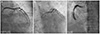
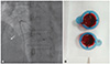
The patient received 300 mg oral aspirin and 600 mg clopidogrel in addition to intravenous unfractionated heparin (bolus 5000 IU, infusion 80 IU/hr). An emergency angiography was performed via the right radial artery with a 5 Fr Judkins catheter (Vista Brite Tip, Cordis, Miami, FL, USA). The left coronary angiography showed minimal atherosclerotic changes in the mid segment of the left anterior descending artery without visualization of the LCX (Fig. 1A and B). There were no collateral flows to the LCX. The right coronary angiography revealed a relatively large RCA and a total thrombotic occlusion in the mid segment (Fig. 1C). We thought that the RCA was the culprit vessel, and a 6 Fr Judkins right (JR) guide catheter was engaged to perform percutaneous coronary intervention. We were concerned about the massive intracoronary thrombosis because, due to the poor left ventricular systolic function, the absence of the reflow phenomenon or thrombus propagation to the distal coronary artery after revascularization could be fatal. A thrombus aspiration was attempted with a coronary thrombosuction device (Thrombuster II, Kaneka Medix, Kaneka, Japan), but it was not effective because of the extremely large thrombus burden. We decided to extract the massive thrombus directly using the 6 Fr JR guide catheter instead of the thrombosuction device because the diameter of the RCA was large enough for a 6 Fr guide catheter to advance through the lumen. The JR catheter was deeply positioned into the mid segment, and thrombus aspiration was performed carefully several times (Fig. 2A). After these procedures, the system was removed from the sheath and flushed. The remnant thrombus was aspirated using a 5 Fr daughter catheter (Heartrail, Terumo, Tokyo, Japan) and a coronary thrombosuction device. The massive fresh red thrombus was extracted (Fig. 2B) and the final coronary angiography revealed the complete recovery of coronary flow. The super-dominant RCA, with its posterolateral branches supplying the lateral wall of the left ventricle corresponding of the territory of the LCX appeared (Fig. 3A). The patient subsequently received intracoronary abciximab (250 µg/kg bolus, 10 µg/min for 12 hours). Dual antiplatelet therapy, including aspirin and clopidogrel, and intravenous unfractionated heparin were continued for eight days.
A coronary angiography was repeated on day 8. A minimal remnant thrombus was observed in the large RCA, but not in the distal branches (Fig. 3B). No significant luminal stenosis was observed, and this was confirmed by intravascular ultrasonography (Atlantis SR Pro, Boston Scientific, Natick, MA, USA) (Fig. 3C, D, and E). The patient's clinical condition stabilized without any complications and she was discharged with aspirin and clopidogrel five days later.
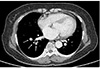
Two weeks after discharge, the patient complained of chest discomfort and dyspnea on exertion. There were no remarkable changes in cardiac enzymes, or on her EKG and echocardiography. However, multiple pulmonary thromboemboli were noted on the chest CT scan (Fig. 4). The patient was treated with warfarin for six months after discontinuation of dual antiplatelets. After the six months chest CT scan was repeated and there was no evidence of pulmonary thromboembolism.
Discussion
The absence of the LCX is an extremely rare congenital anomaly with a frequency of only 0.003% in 126595 patients who underwent coronary angiography.1) The condition is commonly compensated for with an oversized super-dominant RCA that extends into the LCX territories to minimize the area of hypoperfusion. Due to the compensation mechanism, this vascular anomaly is usually considered benign. However, when acute occlusion of the RCA occurs, it is usually equivalent to a two-vessel coronary artery disease involving both the RCA and LCX. This occlusion results in inferior, posterior, and lateral wall myocardial infarction.
Giant coronary artery diseases, such as coronary artery ectasia, may increase the risk of ischemic symptoms or myocardial infarctions with or without an occlusion of the coronary artery caused by sluggish coronary flow or turbulent flow. Complications from this disease usually occur as thromboembolic phenomena primarily due to thrombosis in the ectasic segment of the coronary artery.6)
In the present case, the LCX was not seen on the left coronary angiography and the huge RCA was occluded on the right coronary angiography. Though multidetector-row computed tomography could provide much more information about coronary anomalies and it might be the best modality for confirmation, the patient did not want to underrgo further evaluation.7) However, many distal RCA branches were noted on the coronary angiography after revascularization, which extended to the lateral wall of the left ventricle without collateral flow from the RCA to the LCX. No anomalous vessel arising from aorta was observed on the chest CT scan, which was taken for the diagnosis of pulmonary thromboembolism. These results demonstrate the absence of the LCX.
According to Yip et al.8) an infarct-related artery (IRA) with a reference lumen diameter (RLD) lager than 4 mm and high-burden thrombus formation results in a significantly higher incidence of unsuccessful reperfusion than an IRA with a RLD <4 mm in patients with AMI. Slow and turbulent flow in a large diameter coronary artery is associated with thrombus formation and may initiate platelet activity and damage the endothelium, which causes formation of a massive thrombus without significant coronary artery stenosis.9)
In cases of IRAs with massive thrombus burden, the current reperfusion techniques are not effective. It is important to extract a thrombus completely to restore the coronary flow and to prevent the no-reflow phenomenon. Onoda et al.10) previously extracted a massive intracoronary thrombus using a 6 Fr JR catheter and demonstrated that the procedure was safe and effective. They inserted a 6 Fr JR catheter into an 8 Fr Amplatz catheter using the 'mother and daughter' technique, and the mother catheter was shortened so the daughter catheter could reach the distal segment of the RCA.
In this case, we extracted most of the thrombus directly using a 6 Fr JR catheter via the right radial approach. Our strategy required less time. The JR catheter was positioned deeply using careful manipulation with a clock-wise rotation and gentle movements without causing major injury to the vessel wall. Possible complications during aggressive mechanical thrombectomy procedures include a collapse of the coronary artery due to strong aspiration or formation of a secondary embolism caused by the extracted thrombus falling into the coronary artery or aorta.11) This technique should be considered only in selected cases when an operator is an expert in the transradial procedure and the coronary artery is large enough for the procedure.
The mechanism of massive red thrombus formation in this case was not clear, but the patient's thrombogenic condition might have been due to her advanced malignancy stage. The patient had stage IV lung cancer and was undergoing her sixth cycle of pemetrexed and cisplatin. The contribution of an intra-coronary glycoprotein IIb/IIIa inhibitor to dissolve the clot remains unclear. According to Fujii et al.12) an intracoronary thrombus consisting of red blood cells is a high-burden thrombus, while those consisting of platelets are low-burden thrombi. Glycoprotein IIb/IIIa inhibitors do not have primary fibrinolytic activity. However, inhibitors can help dissolve the remnant intracoronary thrombus by assisting spontaneous fibrinolysis especially in an active thrombogenic state. Hypercoagulable studies at admission including protein C and S, anti-thrombin III, Factor V Leiden, prothrombin, homocysteine, lupus anticoagulant, and antiphospholipid Ab were all within normal limits or negative in our patient. Consequently, a thrombogenic medical condition, such as malignancy, may cause pulmonary thromboembolism and myocardial infarction of the large coronary artery despite the continuous use of dual antiplatelets.
In conclusion, we have described an effective mechanical aspiration technique, which may become an alternative procedure in cases of massive thrombi situated in large diameter coronary arteries in order to avoid the slow flow or no-flow phenomenon, and to achieve complete revascularization.

 XML Download