

 PDF
PDF ePub
ePub Citation
 Print
Print


Introduction
The histologic confirmation is necessary for the definite diagnosis and is essential in determining the treatment for patients with cardiac tumors. Depending on the various clinical situations, tissue diagnosis can be made with less invasive methods including cytologic evaluation of pericardial or pleural fluids, or percutaneous biopsy of metastatic subcutaneous lymph nodes. Open heart surgery, mediastinoscopy, and metastatic mass exploration are more invasive methods that require general anesthesia. Percutaneous transvenous biopsy of cardiac tumor can be done under the guidance of transesophageal echocardiography (TEE). However, general anesthesia is also needed for this procedure.
Recently, intracardiac echocardiography (ICE) has been introduced to assist many cardiac procedures and it provides high-quality imaging like TEE. Although it needs local anesthesia and venous puncture with relatively large catheter, it can give us a good guidance during the biopsy of cardiac tumors. In this report, we present two cases of cardiac masses documented by percutaneous transvenous biopsy under the guidance of an ICE.
Cases
Case 1
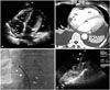
A 51-year-old woman was admitted to our hospital with a 3-day history of progressive dyspnea and generalized edema. The initial electrocardiogram revealed atrial fibrillation with rapid ventricular response, low QRS voltage, and poor R progression. The chest X-ray showed a water-bottle shaped cardiomegaly and bilateral pleural effusion. The transthoracic echocardiogram (TTE) showed a large amount of pericardial effusion with diastolic right atrial (RA) and ventricular collapse associated with inferior vena caval plethora. It revealed infiltrative masses along the superior and posterior aspect of RA wall (Fig. 1A). Clinical symptoms improved after pericardial drainage of serosanguinous fluid. The computed tomographic (CT) scan revealed infiltrating mass on RA wall (Fig. 1B) and multiple tiny nodules scattered in the whole lung fields.
Because the cytologic examination from the pericardial fluid was inconclusive, we performed percutaneous biopsy of the RA mass in order to establish a pathological diagnosis. After placement of an 8 Fr Ultra ICE catheter (AcuNav, Siemens Medical Solution, Mountain View, CA, USA) into the RA, a 6 Fr biopsy catheter was inserted into RA and several pieces of RA mass were obtained (Fig. 1C and D). The procedure was well tolerated under local anesthesia, and no complication occurred during the procedure. The pathologic diagnosis was infiltrating angiosarcoma. Although the patient was treated with systemic chemotherapy, she died from hepatic rupture associated with multiple liver metastases at 18 months.
Case 2
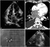
A 53-year-old man visited our hospital with a 10-day history of progressive dyspnea and weight loss of 4 kg. The initial electrocardiogram revealed normal sinus rhythm with poor R progression. The chest X-ray showed large pleural effusion at the right lung field. The TTE demonstrated a large infiltrating mass on both atrial walls (Fig. 2A). The CT scan revealed a huge mass infiltrating both atrial wall and posterior mediastinal extension (Fig. 2B).
Because cytologic confirmation of the pleural fluid was negative and percutaneous needle biopsy of the mediastinal mass could be a high-risk procedure, we performed percutaneous RA mass biopsy in order to get prompt specimens. A 6 Fr cardiac biopsy catheter and an 8 Fr Ultra ICE catheter were percutaneously inserted into the RA through the right and left femoral vein, respectively. ICE demonstrated the tumor spreading at the posterior aspect of the RA wall. We obtained several pieces of RA mass with the ICE monitoring (Fig. 2C and D). There was no complication associated with the procedure. The patient was diagnosed with small cell lung cancer with metastasis to the RA. The patient died from disease progression at 10 months, despite of systemic chemotherapy.
Discussion
The malignant primary cardiac tumors are extremely uncommon, whereas metastatic cardiac tumors occur more frequently.1,2,3) In a patient suspected of malignant cardiac tumor without an option for curative surgical treatment, histopathological confirmation is a mandatory option next to treatment strategy, regardless of its primary focus. Depending on the various clinical presentations, pathologic diagnosis can be made with less invasive methods such as cytologic evaluation of pericardial or pleural fluids, or percutaneous biopsy of metastatic subcutaneous lymph node. More invasive methods of tumor biopsy through open heart surgery, mediastinoscopy, or metastatic mass exploration may be needed when less invasive approach is not confirmative. Percutaneous transvenous biopsy of cardiac tumor is a good alternative using less invasive tool for tissue diagnosis.
Traditionally, the fluoroscopy guidance is the most commonly used for the blunt endomyocardial biopsy. Because the fluoroscopy is inadequate to target the mass lesion, especially in patients with cardiac tumors, many researchers use TTE or TEE to guide the prompt site for biopsy.4)5) However, TTE gives less clear cardiac images than TEE, especially for the posterior wall mass and the patient with poor echo window,6) and TEE requires general anesthesia during the procedure.
Recently, the ICE has been introduced to guide several cardiac procedures including device closure for congenital shunts, septal puncture, radiofrequency catheter ablation for arrhythmias, and intracardiac biopsy of tumor.7,8,9,10,11) Because ICE can give us clearer cardiac imaging without using the general anesthesia, it is more practical and patient-friendly. It can also reduce radiation exposure for the echocardiographer during the procedure.
In this report, we showed the feasibility of the ICE during targeting of the biopsy catheter, without a requirement of patient sedation. The use of ICE during the procedures increase diagnostic yield and can reduce the incidence of cardiac complications associated with inadequate targeting of the biopsy catheters. The indication of ICE guided biopsy has not been established yet. It can be used for the biopsy of the mass from right-side of the heart, especially in patients with high risk for anesthesia.9,10,11) These ICE-guided techniques allow the physician to safely and effectively perform endocardial biopsy with short total procedure time and less exposure to radiation.
 XML Download