

 PDF
PDF ePub
ePub Citation
 Print
Print


Introduction
Aneurysms of the sinus of Valsalva are rare cardiac anomalies that may be congenital or, in rare cases, acquired, and most commonly involve the right coronary sinus (90%).1)2)3)4) Congenital aneurysms are often caused by weakness at the junction of the aortic media and the annulus fibrosus.3) The acquired forms are caused by infection, degenerative disease or thoracic trauma. Although the majority of unruptured aneurysms do not produce symptoms of cardiac dysfunction until aneurysm rupture occurs, there have been a few reported cases in which patients with an unruptured aneurysm of the sinus of Valsalva presented with exertional dyspnea due to right ventricular outflow tract (RVOT) obstruction.5)6) We report the case of a patient who presented with exertional dyspnea and was found to have an unruptured aneurysm of the sinus of Valsalva causing significant RVOT obstruction.
Case
A 66-year-old man with a medical history of hypertension and type 2 diabetes presented at our cardiology clinic with a 3-year history of worsening dyspnea on exertion (New York Heart Association class I-II). On admission, blood pressure was 132/62 mm Hg, pulse rate was 80/min, body temperature was 36.7℃ and respiratory rate was 22/min. A systolic ejection murmur, grade III/IV, was heard at the second and third intercostal spaces along the left sternal border. The rest of the physical examination was normal. Plain chest X-ray revealed normal chest findings. Electrocardiography demonstrated a normal sinus rhythm with left ventricular hypertrophy.
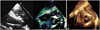
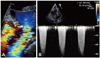
Coronary angiography indicated that there was no significant stenosis of the coronary artery. B-type natriuretic peptide was 450 ng/mL. Laboratory findings were unremarkable except for mild proteinuria on urinalysis. On transthoracic echocardiography, the left ventricular ejection fraction was 55% with normal wall motion, and mild diastolic dysfunction was present (E/A ratio 0.8 deceleration time 250 msec). Transthoracic two-dimensional (2D) and three-dimensional (3D) echocardiography revealed a 1.35×3.0 cm unruptured aneurysm of the right sinus of Valsalva protruding into the RVOT (Fig. 1) with mild aortic regurgitation. Color Doppler echocardiography showed turbulent flow acceleration around the aneurysm, but did not indicate left to right shunt flow (Fig. 2A). Continuous wave Doppler demonstrated that the peak velocity at the level of the aneurysm was 3.38 m/s, and the calculated peak pressure gradient was 46 mm Hg (Fig. 2B).
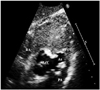
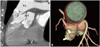
As mentioned above, transesophageal echocardiography revealed aneurysm; however, further cardiac abnormalities were not identified. On contrast echocardiography, subcostal images demonstrated a significant border between the body of the aneurysm and the chamber of the right ventricle upon administration of intravenous echo contrast (Fig. 3). 64-slice multidetector computed tomography (MDCT) showed the presence of an eccentric, lobulated, 3.5 cm aneurysm protruding into the RVOT, with the upper lobulated part located just below the pulmonary valve and the digital-shaped lower part obstructing the RVOT (Fig. 4).
Discussion
Aneurysms of the sinus of Valsalva are rare cardiac abnormalities that may be congenital or acquired. Congenital aneurysms, which are more common than the acquired varieties, slowly progress without symptoms of cardiac dysfunction until aneurysm rupture occurs. However, in rare cases, unruptured congenital aneurysms may produce symptoms of cardiac dysfunction due to RVOT obstruction. In our case, the patient was asymptomatic during his adolescence and most of his adulthood and had no history of risk factors for acquired aneurysm. Moreover, he had experienced progressively-worsening exertional dyspnea over the course of 3 years. Therefore, we regarded the etiology of his condition to be a congenital unruptured aneurysm of the right sinus of Valsalva. We then came to the conclusion that his symptoms may have been due to the slowly progressive nature of congenital aneurysms, which eventually led to significant RVOT obstruction.
In this case, the patient was diagnosed with an unruptured aneurysm of the sinus of Valsalva using transthoracic 2D echocardiography, transthoracic 3D echocardiography, transesophageal echocardiography, contrast echocardiography and 64-slice MDCT. Al-though transthoracic 2D echocardiography is the initial diagnostic tool typically used to detect aneurysms of the sinus of Valsalva4)7) and is sufficient to confirm diagnosis, in the present case, it was difficult to determine the shape of the aneurysm and neighboring st-ructures. Therefore, 64-slice MDCT and real-time 3D echocardiography were used to evaluate the anatomy of this aneurysm and to demonstrate the mechanism of RVOT obstruction. In addition, contrast echocardiography is a useful tool for detecting aneurysms through differentiation of the border between the body of aneurysm and the chamber of the right ventricle.
For patients with unruptured aneurysms of the sinus of Valsalva, symptoms of cardiac dysfunction are important indications for tr-eatment.2)3)8) However, whether asymptomatic patients with unruptured aneurysms require surgical intervention remains a controversial issue. Nevertheless, if unruptured aneurysms show progressive enlargement on serial evaluation, surgical intervention is needed.
This patient was found to have an unruptured aneurysm arising from the right coronary sinus. We determined that his exertional dyspnea was attributable to RVOT obstruction by an unruptured aneurysm of the right sinus of Valsalva, as all other causes were ruled out. Surgical repair could be an appropriate treatment for aneurysms exhibiting progressive enlargement and the potential for rupture.
 XML Download