

 PDF
PDF ePub
ePub Citation
 Print
Print


Introduction
In a differential diagnosis of a wide complex tachycardia (WCT), prior to which a 12-lead surface electrocardiogram (ECG) was taken, a comparison of QRS morphology between the sinus rhythm and the WCT is helpful. An identical QRS morphology between sinus rhythm and WCT strongly suggests supraventricular tachycardia (SVT) whereas a contralateral bundle branch block (BBB) morphology during tachycardia with a preexisting BBB strongly suggests ventricular tachycardia (VT).1) Contrary to this rule, we present a case of SVT with left BBB (LBBB) aberration but right BBB (RBBB) during sinus rhythm.
Case
A 45-year-old woman with a 30-year history of palpitations presented herself for an electrophysiologic (EP) study and a catheter ablation. Her present arrhythmia was a LBBB morphology tachycardia (LBBB-T) at 200 beats/min which was converted into a sinus rhythm with intravenous adenosine. During the EP study, multipolar catheters were placed in the high right atrium (HRA), His-bundle region (His), right ventricle (RV), and coronary sinus (CS).
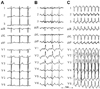
At the beginning of the EP study, the surface ECG did not show any BBB or intraventricular conduction delays (Fig. 1A), and the baseline conduction intervals were within normal limits. The AH interval was 72 msec, and the HV interval was 54 msec. However, an RBBB occurred due to mechanical trauma during His-bundle catheter placement (Fig. 1B). The baseline EP study showed ventriculo-atrial (VA) conduction was present through both the atrioventricular (AV) node and the concealed accessory pathway (AP) in the left posterior, which was blocked by 250 msec pacing with an AV Wenckebach cycle length of 310 msec. The clinical LBBB-T (cycle length: 330-350 msec) was easily induced by catheter-manipulation and spontaneous premature atrial complexes (Fig. 1C). LBBB-T has several possible mechanisms including orthodromic atrioventricular reentrant tachycardia (ORT) with an LBBB aberration using a concealed AP, antidromic atrioventricular reentrant tachycardia (ART) using various types of right-sided APs anterogradely, RV VT, and bundle branch reentry VT.
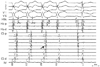
Fig. 2 shows that the LBBB-T terminated spontaneously and was followed by a sinus beat with a RBBB.
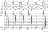
The LBBB-T terminated without any following atrial activation; therefore, VT with 1:1 retrograde conduction was ruled out. The His-bundle activation during the tachycardia preceded the beginning of the QRS complex, which ruled out an ART. The site of the earliest atrial activation during the LBBB-T was in the mid-CS rather than in the His-bundle. Delivery of premature ventricular stimulus during the His-bundle refractory terminated tachycardia without atrial activation. This confirmed that the tachycardia was an ORT using a concealed left posterior AP. Ablation was successful and the tachycardia could no longer be induced. After ablation, VA conduction was present through the AV node only, but a VA conduction block occurred at the VH level with a Wenckebach pattern (Fig. 3), which suggested a conduction abnormality in the His-Purkinje system or between the His-Purkinje system and the ventricle.
Discussion
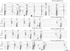
Bundle branch block aberrancy during SVT in the presence of pre-existent contralateral BBB is extremely rare, even in the presence of a His-Purkinje system dysfunction. A functional or anatomical block at one or more levels in the conduction system can cause LBBB.2) It can occur at the His bundle level (dedicated fibers to LB) before its bifurcation, the left bundle branch level, the left fascicle level (due to variation in the anatomy of fascicles3)), or diffuse disease of the very distal ramifications of the left bundle. Fig. 4 shows conduction patterns during programmed and burst stimulation from the HRA. Burst pacing at 280 msec revealed a conduction delay between the His and BB resulting in an infra/intra Hisian block (Wenckebach type). The interval following a non-captured beat allowed recovery of the left bundle, and this resulted in the re-appearance of the RBBB QRS morphology similar to that of baseline conduction. Early-coupled atrial extrastimuli caused a progressive delay within the His bundle or between the His and BB, and the RB potential to the QRS interval became longer and fixed during the LBBB morphology. The lower tracing in Fig. 4 showed that the LBBB did not recover from previous stimulation, but the interval from His to RB became longer by a premature stimulation without a change in LBBB morphology. This implies that a certain amount of conduction delay between the His and bundle branch may be the cause of manifestation of LBBB in this case.
We propose that conduction delay between His and bundle branch may be the possible mechanism of the LBBB aberration with SVT during preexisting RBBB.
 XML Download