

 PDF
PDF ePub
ePub Citation
 Print
Print


Introduction
Vascular smooth muscle cells (VSMCs) are major cells in the neointimal region and play a role in vascular diseases.1) Unlikely other cells, VSMCs can switch phenotypes between contractile (differentiation) and proliferative (de-differentiation) state in response to various pathological stimuli. This vascular phenotype switching contributes to the development of cardiovascular diseases such as atherosclerosis, restenosis, and hypertension.2) Therefore, regulating the VSMC phenotype may have potential as a therapeutic target for treating pathological vascular diseases.
The most significant breakthrough in modulating gene expression is the discovery of microRNAs. MicroRNAs (miRs) are small, non-coding RNAs that negatively regulate gene expression by degradation or translational inhibition of microRNA target genes.3)4) MicroRNAs are currently considered key regulators or possible biomarkers of various pathological diseases, ranging from various cancers to cardiovascular diseases. They are expressed in a cell- or tissue-specific manner and play important roles in many cellular processes such as cell differentiation, proliferation/growth, apoptosis, and migration. Recent studies have shown that microRNAs are phenotypic regulators of VSMCs. For example, miR-663, miR-221, miR-143, miR-145, miR-133, and miR-26 regulate VSMC differentiation and phenotypic changes.5)6)7)8)9)10) Our group has studied the roles of microRNAs in VSMCs as well as in a rat carotid injury model. We reported that miR-132 induces VSMC differentiation and that miR-132 delivery reduces neointimal hyperplasia.11) Several studies have revealed that miR-18a-5p is involved in development,12) proliferation,13) and differentiation.14) However, the role of miR-18a-5p in VSMC proliferation and differentiation is currently unknown.
In the present study, we identified new microRNAs that regulate VSMC differentiation in a rat carotid artery balloon injury model. We also demonstrated that miR-18a-5p promotes VSMC differentiation by downregulating syndecan4.
Materials and Methods
Reagents, miR-18a-5p mimic, and antibodies
Human recombinant platelet derived growth factor-BB (PDGF-BB) was purchased from Sigma (St. Louis, MO, USA). MiRNA-18a-5p and the miRNA control were purchased from Bioneer Co. (Daejeon, Korea).
Gapdh (sc-32233), syndecan4 (sc-15350), smooth muscle (SM) 22α (sc-373928), and SM α-actin (sc-130617) were purchased from Santa Cruz Biotechnology (Santa Cruz, CA, USA). Smad2 (#3103) was purchased from Cell Signaling Technology (Danvers, MA, USA).
Rat carotid injury model and microRNA microarray
Rat carotid injury experiments were performed as described previously. Briefly, 8-week-old male Sprague-Dawley rats (n=8, Damul, Daejeon, Korea) were used, and all experimental procedures were approved by the Chonnam National University Medical School Research Institutional Animal Care and Use Committee. Rats were anesthetized for balloon injury, and the right common carotid artery was exposed by a midline cervical incision. A 2 Fr Fogarty balloon catheter was placed at the puncture site of the external carotid artery and retracted three times under mild balloon inflation pressure to denude the endothelium. The left carotid artery was used as the sham control. The arteries were harvested at 1, 7, and 14 days after injury. microRNA expression profiles were analyzed with an Agilent microRNA microarray (Agilent Technologies, Palo Alto, CA, USA).
Cell cultures
Vascular smooth muscle cells were isolated from the aortic media of male rats by enzymatic dissociation as described previously.15) VSMCs were used at passages 5-11. A10 cells were obtained from the American Type Culture Collection (Manassas, VA, USA). The cells were grown in Dulbecco's modified Eagle's medium (DMEM) with 10% fetal bovine serum (FBS).
Differentiation and de-differentiation of vascular smooth muscle cells
Vascular smooth muscle cell differentiation was induced by serum deprivation. Briefly, cells were seeded into 6-well plates at 50% confluence and maintained in 0.5% DMEM media containing 0.5% FBS for 4 days. Cells were serum-starved for 24 hours and treated with PDGF-BB (25 ng/mL) for 3 days to de-differentiate.
miR-18a-5p mimic
Double-stranded RNAs were designed to overexpress the endogenous mature miR-18a-5p. Primary VSMCs were transfected with miR-18a-5p mimic or negative control miRNA (75 nM, Bioneer) using the RNAiMax reagent (Invitrogen, Carlsbad, CA, USA) according to the manufacturer's protocol.
Plasmid construction and RNA interference
pCMV-SPORT6-Smad2 was obtained from KUGI (Seoul, Korea). The pGL3-441 SM22α promoter-luciferase construct was kindly provided by Prof. Michael S. Parmacek (University of Pennsylvania, Philadelphia, PA, USA). The pcDNA-syndecan4 full wild construct was kindly provided by Prof. Eok-Soo Oh (Ewha Womans University, Seoul, Korea). All plasmids were confirmed by sequencing.
Syndecan4 knockdown was performed using its siRNA (si-syndecan4, 50 nM, cat no. sc-270178, Santa Cruz Biotechnology, Santa Cruz, CA, USA). An siRNA targeting rat syndecan4 was transfected into A10 cells using the RNAiMax reagent. Nontargeting siRNA (Bioneer) was used as a negative control.
Luciferase assay
Cells (A10 and 293T) were seeded in 24-well plates 18 hours before transfection. The pGL3-441 SM22α promoter and pCMV-β-galactosidase were transfected with Lipofectamine Plus reagent (Invitrogen, Carlsbad, CA, USA) or PolyFect reagent (Qiagen, Valencia, CA, USA), according to the manufacturer's instructions. The cells were harvested 48 hours after transfection, and luciferase activity was measured and normalized against β-galactosidase activity as an internal control.
Western blots
Cells were harvested with RIPA lysis buffer as described previously.15) Cell extracts were subjected to sodium dodecyl sulfate-polyacrylamide gel electrophoresis and transferred to a PVDF membrane. Western blotting was performed using anti-SM α-actin, anti-SM22α, anti-syndecan4, anti-Smad2, and anti-GAPDH. The blots were developed using Immobilon Western Detection Reagents (Millipore, Billerica, MA, USA).
Quantitative real time polymerase chain reaction
Total RNAs were extracted with a Trizol kit according to the manufacturer's instructions.
Real time polymerase chain reaction (RT-PCR) was performed as described previously.16) Polymerase chain reaction (PCR) was performed using the following oligonucleotide primers: for SM α-actin, sense, 5'-AGTCGCCATCAGGAACCTCGAGAA-3', and antisense, 5'-GC CAGATCTTTTCCATGTCGTCCC-3'; for SM22α, sense, 5'-CCCACAAAC GACCAAGCCTTTTCT-3', and antisense, 5'-CCTGTTCCATCTGCT GAAGACCA-3'; for calponin, sense, 5'-ACAACACCCAAAGGAAGCAC-3', and antisense, 5'-TCACTGCAAAACCAAACTGC-3'; for Smad2, sense, 5'-CCAGGTCTCTTGATGGTCGT-3', and antisense, 5'-ACTGGTGTCTC CACCCTCTG-3'; for syndecan4, sense, 5'-GATAACCACATCCCC GAGAA-3', and antisense, 5'-CACAATCAGAGCTGCCAAGA-3'; for glyceraldehyde-3-phosphate dehydrogenase, sense, 5'-AACCCAT CACCATCTTCCAGGAGC-3', and antisense, 5'-ATGGACTGTGGTCAT GAGCCCTTC-3';
miR-18a-5p quantitative real time polymerase chain reaction
RNAs from rat carotid arteries and VSMCs were isolated with TRIzol (Invitrogen, Carlsbad, CA, USA) to detect mature miR-18a-5p levels. qRT-PCR for miR-18a-5p was performed using cDNA generated from 10 ng of total RNA using the TaqMan MicroRNA Reverse Transcription kit (part no. 4366596, Applied Biosystems, Foster City, CA, USA). 18S was used as an internal control. Quantification of the mRNA amounts was done with SYBR Green PCR kit (Applied Biosystems, Foster, CA, USA).
miRNA target prediction
We used the miR Base Target database to find putative miR-18a-5p targets (www.mirbase.org). Candidate target genes were determined by qRT-PCR.
Results
miR-18a-5p mimic increases vascular smooth muscle cell differentiation
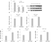
To explore the role of miR-18a-5p in the rat carotid injury model and VSMCs, we first investigated miR-18a-5p expression in carotid arteries at 1, 7, and 14 days after injury. Using microRNA qRT-PCR analysis, we observed induction of miR-18a-5p 1 day after carotid injury compared with the sham control (Fig. 1A). Next, we evaluated whether miR-18a-5p affected proliferation or differentiation of VSMCs. MiR-18a-5p mimic did not affect proliferation of VSMCs (data not shown). As shown in Fig. 1B, C, and D, the miR-18a-5p mimic increased SMα-actin and SM22α protein amounts. We further determined other VSMC differentiation markers using qRT-PCR. SM α-actin, SM 22α, and calponin mRNA levels were induced by transfection of the miR-18a-5p mimic into VSMCs (Fig. 1E, F, and G).
miR-18a-5p expression during differentiation and de-differentiation of vascular smooth muscle cells
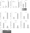
Unexpectedly, the qRT-PCR analysis revealed enhanced SM22α and SMα-actin mRNA levels in injured arteries compared with those in sham-operated arteries (Fig. 2A and B). We determined the expression of miR-18a-5p and differentiation markers during differentiation and de-differentiation of VSMCs to verify whether miR-18a-5p was involved in the VSMC phenotype. We found that SM22α protein expression was induced in VSMC differentiation medium (Fig. 2C). SM α-actin and SM22α mRNA levels increased in differentiated VSMCs (Fig. 2D and E). As shown in Fig. 2F, miR-18a-5p expression increased after exposure to differentiation medium. De-differentiated VSMCs induced by treatment with PDGF-BB showed decreased SM22α and SM α-actin mRNA levels. In addition, SM α-actin protein expression decreased in PDGF-BB-treated VSMCs (Fig. 2I). Interestingly, we found that miR-18a-5p expression was downregulated in PDGF-BB-stimulated VSMCs (Fig. 2J).
Syndecan4 is a target gene of miR-18a-5p in vascular smooth muscle cells
We used bioinformatics tools to identify candidate miR-18a-5p target genes. Considering that miR-18a-5p is involved in VSMC differentiation, we hypothesized that downstream miR-18a-5p target genes might be associated with differentiation. Therefore, we selected eight genes (NFAT5, Hiflα, Hsf2, Tmx4, CTGF, KLF6, syndecan4, and Smad2). These candidate genes have been reported to relate to differentiation.
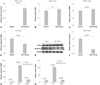
We first evaluated whether miR-18a-5p expression was induced by transfection of the miR-18a-5p mimic into VSMCs (Fig. 3A). Next, we tested whether miR-18a-5p downregulated expression of the candidate target genes. Although Smad2 contains the miR-18a-5p binding site in the Smad2 3' untranslated region, Smad2 protein levels remained unchanged in miR-18a-5p-transfected VSMCs (Fig. 3B). This result indicates that Smad2 is not a direct target of miR-18a-5p. Of the genes examined, syndecan4 mRNA levels decreased significantly in both VSMCs and A10 cells transfected with miR-18a-5p (Fig. 3C and D). In addition, syndecan4 protein expression was clearly reduced in VSMCs transfected with miR-18a-5p (Fig. 3E). Quantification of syndecan4 protein levels is shown in Fig. 3F. To further identify whether syndecan4 was a target of miR-18a-5p in balloon-injured rat carotid artery, we examined syndecan4 mRNA expression levels by qRT-PCR. As shown in Fig. 3G, syndecan4 mRNA was highly expressed 7 days after balloon injury. In addition, we found that Smad2 expression was enhanced 7 days after carotid injury (Fig. 3H).
Syndecan4 regulates Smad2 expression in vascular smooth muscle cells
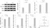
To identify whether syndecan4 is involved in VSMC differentiation, we examined differentiation marker genes in VSMCs. We confirmed that the amounts of syndecan4 mRNA and protein largely increased by transfection with the syndecan4 expression vector in A10 cells (Fig. 4A and E). However, the SM differentiation markers SM22α and SM α-actin remained unchanged by overexpressing syndecan4 in A10 cells (Fig. 4B and C). Unexpectedly, we found that Smad2 protein expression decreased in syndecan4-overexpressing cells (Fig. 4D and F). To further determine regulation of Smad2 by syndecan4, the cells were transfected with VSMCs with syndecan4 siRNA. Endogenous syndecan4 decreased by syndecan4 siRNA treatment (Fig. 4G and H). Despite the significant reduction in syndecan4, syndecan4 siRNA did not affect the smooth muscle differentiation markers (Fig. 4G). However, Smad2 expression increased slightly by syndecan4 siRNA (Fig. 4I).
Smad2 promotes vascular smooth muscle cell differentiation
Although the miRBase prediction database showed several miR-18a-5p target genes, Smad2 did not decrease following transfection with the miR-18a-5p mimic. Because Smad2 was downregulated by syndecan4 in VSMCs, we sought to determine whether Smad2 was involved in differentiation. We transiently transfected pCMV-SPORT6-Smad2 or empty vector into A10 cells and examined Smad2 protein expression. When cells were transfected with Smad2, SM α-actin and SM22α protein expression increased (Fig. 5A-D). SM 22α is a differentiation marker. Cells were transfected with the -441 SM22α minimal promoter construct in the presence or absence of Smad2. As shown in Fig. 5E and F, exogenous Smad2 significantly activated the SM22α promoter in both A10 and 293T cells.
Discussion
We showed that miR-18a-5p was upregulated at the early phase after rat carotid balloon injury and promoted VSMC differentiation. The main observation of our study is that miR-18a-5p promoted VSMC differentiation by inhibiting syndecan4 expression. Although syndecan4 did not directly affect VSMC differentiation, miR-18a-5p-repressed syndecan4 induced Smad2, which activated VSMC differentiation (Fig. 5G). The detailed molecular mechanism needs to be clarified in a future study.
We demonstrated that miR-18a-5p promoted the expression of VSMC differentiation marker genes (i.e., SM α-actin and SM22α). In addition, we found that miR-18a-5p expression was upregulated at the early time point after balloon injury. We further demonstrated that miR-18a-5p expression increased during VSMC differentiation, whereas it decreased in VSMC de-differentiation medium. Therefore, we suggest that miR-18a-5p is a novel modulator of the VSMC phenotypic switch.
Considering the effect of VSMC differentiation by miR-18a-5p, we hypothesized that miR-18a-5p must downregulate anti-differentiation genes. Among several genes, we found that syndecan4 mRNA and protein expression were markedly downregulated by miR-18a-5p in VSMCs. However, the downregulation of syndecan4 by overexpression of miR-18a-5p in VSMCs was not completely mimicked in the rat carotid injury model. In our animal experiment, the syndecan4 transcript level was dramatically enhanced in injured arteries 7 days after carotid injury. The reason is attributed to a decrease in miR-18a-5p expression over time after carotid injury.
Syndecan4 is a family of heparan sulfate proteoglycan. Studies have demonstrated that syndecan4 is involved in cardiovascular diseases, such as cardiac hypertrophy and myocardial infarction as well as arterial restenosis after angioplasty.17)18) Herum et al.19) reported that syndecan4 is involved in differentiation from cardiac fibroblasts to myofibroblasts in response to mechanical stress such as pressure overload. However, our results show that transfection of A10 cells with syndecan4 did not affect VSMC differentiation markers. Our results suggest that syndecan4 does not act as an inducer of VSMC differentiation. Syndecan4 could regulate expression of differentiation-associated genes as a possible mechanism of miR-18a-5p-induced VSMC differentiation.
Although syndecan4 is a candidate target of miR-18a-5p, it does not show VSMC differentiation characteristics. Therefore, we investigated whether Smad2 is a downstream gene of syndecan4 in VSMCs. We found that syndecan4 regulates Smad2 expression in VSMCs. Indeed, overexpression of syndecan4 reduced Smad2 protein expression, whereas knockdown of syndecan4 increased expression of Smad2. Xie et al. showed that Smad2 acts as a critical regulator of VSMC differentiation in neural crest cells.20) We found that Smad2 induced protein expression and promoter activity of SM22α in VSMCs.
In conclusion, we identified miR-18a-5p as a novel regulator of VSMC differentiation. Our findings suggest that miR-18a-5p/Smad2 may be a potential therapeutic for treating pathological vascular diseases such as atherosclerosis and in-stent restenosis.

 XML Download