

 PDF
PDF ePub
ePub Citation
 Print
Print


Introduction
A tunnel from aorta to right atrium is a very rare congenital anomaly. This anomaly was first described by Coto et al.1) as the aorta-right atrial communication. In this congenital anatomy, a large vascular link established a shunt originating from the aortic root and terminating in the right atrium. Moreover, this congenital anomaly can be associated with a coronary artery anomaly. We present a case of the aorta-right atrial tunnel (ARAT), which has a large saccular aneusrysmal ending to the right atrium, with the left anterior descending (LAD) and left circumflex (LCX) coronary arteries arising independently from the different proximal parts of the tunnel.
Case
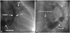
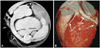
An 18-year-old girl was referred to our institution for the evaluation of a continuous murmur. In her medical history, there were exertional dyspnea and palpitation. Her physical examination showed a grade 3/6 heart murmur, best heard along the upper right sternal border. The chest X-ray revealed that there was no cardiomegaly, but mildly prominent pulmonary vascularity. Electrocardiography was within the normal limits. Upon transthoracic echocardiographic examination (Vivid 7 GE, Norway, 3.5 mHz probe), an abnormal flow within the right atrium which showed a continuous pattern was detected with 2D color Doppler. Other transthoracic echocardiographic findings were within the normal limits. Thereafter, we decided to perform transesophageal echocardiogram for a better delineation of the problem. Transesophageal echocardiography (TEE) confirmed the same flow-pattern in the right atrium. Moreover, there was a suspicious dilated structure within a close vicinity of coronary sinus. We excluded the dilated coronary sinus possibility, because there were no signs in the transthoracic and TEE. Selective left coronary angiography performed by a Judkin's left catheter showed a very large tunnel beginning from the left sinus of Valsalva, coursing posteriorly the aortic root and terminating in the roof of the right atrium with aneursymal dilatations. The tunnel presented two consecutively-different aneursymal dilatations. The distal one was a large saccular aneursym just before entering the RA through a very stenotic ostium with a forceful blood flow, whereas the upper one was in a circular shape. It also revealed that the LAD and LCX coronary arteries arose independently from the different proximal parts of the tunnel: LAD from the upper, and LCX from the lower segment (Fig. 1). However, her right coronary artery was normal and originated from the right sinus of Valsalva. Computerized tomography with 16-slice multi-detector CT scanner CT MX 8000 IDT, Philips Medical Systems) was performed to enlighten the coursing of the tunnel and the locations of ostia of the LAD and LCX. It showed that the tunnel originated from the left sinus of Valsalva, coursed posteriorly and drained into the RA with a narrow opening. It also confirmed the angiography on the origination of the coronary arteries (Fig. 2). All diagnostic modalities showed no other congenital cardiac anomalies. Although she was mildly symptomatic, we offered a surgical ligation because the tunnel was very large and tortuous. The young girl underwent the surgical ligation and a closure. Intraoperative visualization confirmed the findings and diagnosis. First, the tunnel was identified and cut from the right atrial opening of the aneurysmal segment and the proximal segment just behind the LCX of the tunnel. Second, the atrial and distal parts of the tunnel were obliterated through a direct suture. The tunnel was removed surgically and treated with direct closure. Antiplatelet therapy and prophylaxis for infective endocarditis were recommended for 6 months. The patient experienced an uneventful recovery and was asymptomatic with no murmur at the 6-month follow-up.
Discussion
Aorta-right atrial tunnel is a very rare congenital cardiac anomaly, characterized by a vascular tunnel between any one of the aortic sinuses and the right atrium. Etiopathogenesis of this interesting anomaly is not clearly understood. Gajjar et al.2) have proposed that a congenital deficiency of the elastic lamina in the aortic media may cause an extracardiac tunnel from the aorta to right atrium, as from a higher aortic pressure to a lower right atrium pressure. A histologic examination taken from the tunnel has shown that the tissue was similar to the aortic one,3) the findings of which may be pertinent to the proposal of Gajjar et al.2)
In the reported cases, the tunnels originated mostly from the left sinus of Valsalva,2)3)4)5)6) two from the right sinus of Valsalva2)7) and one from the non-coronary sinus of Valsalva.1) In relation to the ascending aorta, the tunnels arising from the left sinus of Valsalva coursed posteriorly, whereas the tunnels originating from the right sinus of Valsalva travelled anteriorly.
The origination of coronary ostia can be different from case to case. In the tunnels from the left sinus, LAD and LCX originated seperately from the left sinus.1)3)4)5)6) In some cases, the left coronary or right coronary artery arose from the proximal part of the ARAT.4)7) In our case, the LAD originated from the proximal part of the tunnel. The LCX also arose from the tunnel, but from the lower part of the tunnel rather than that of the LAD. The origination of coronary ostia is very important for the patients who need surgical closure. In all reported patients, the coronary angiography demonstrated that the distribution of both coronary arteries appeared normal except the sinus node artery. In some cases, no sinus node artery could be identified on the selective coronary angiograms.1)3)4)5) However, no sinus node dysfunction was present in those cases, as in our case. The possible aberrant origin of the sinus node artery should be kept in mind during surgical closure. The visualization of this turtuous and large tunnel originating from one of the aortic sinuses, coursing in the extracardiac way and entering into the RA, can easily differentiate the coronary-cameral fistula and the ruptured aneusrym of the sinus of Valsalva by the echocardiography and aortagraphy. ARAT can also be associated with other congenital anomalies. The most common anomaly is the secundum-type of atrial septal defect (ASD). In a series, 3 of 9 patients had ASD.2) Only in one case, the absent right superior vena cava and the presence of a large left superior vena cava was reported.1) Our case was not associated with other congenital anomalies.
The need for closure remains controversial becasue of its asymptomatic nature. Some patients can suffer from dyspnea and palpitation, since ARAT is a kind of arterio-venous shunt. Authors point out that the closure of an ARAT seems logical, because the closure minimizes the risks for the volume-overload of both ventricles, aneusrysm formation, infective endocarditis and the likelihood of a spontaneous rupture.3) There are several types of management options depending on the type of coronary ostia, coursing and location of the coronary ostia. Most reported cases have undergone surgical closure of the tunnel. However, in the literature, there are several case reports describing the percutaneous transcatheter closure of ARAT with coils, vascular plugs and duct occluders. The catheter-closure method using coil embolization or vascular plug device can be advised if the coronary ostia origins independently from any of the coronary sinuses, the opening of the right atrial end is small and if there is no associated cardiac anomaly.2)4)6)8) Although our patient was mildly symptomatic, we offered the surgical closure because of the large tortuous aneursymal dilatations and the origins of coronary ostia from the tunnel. The tunnel was removed surgically and treated with direct closure.
In summary, ARAT should be included in the differential diagnosis of continuous murmur. Aortagraphy and selective angiography is the essential aproach for the visualization of the relationship with the coronary arteries, its coursing and draining into RA. Any closure options should be recommended to the patients soon after the diagnosis, considering the potential risks for the patients.
 XML Download