

 PDF
PDF ePub
ePub Citation
 Print
Print


Introduction
Atrial septal defect (ASD) is a tissue defect which allows blood passing between both atria, and accounts for approximately 5-10% of all congenital heart defects.1) Under clinical situations such as high proportions of left-to-right shunts, right ventricular volume overloads and paradoxical embolisms, the closure of ASD may be required. For the treatment of ASD, depending on the type and anatomical characteristics, percutaneous closure or surgical repair techniques are applied. The surgical approach is based on closing the defects with a direct suture or patch.2) Embolic complications can occur after surgical interventions,3) but cases reported after primary sutures are uncommon.4)5) Herein, we report two cases of large right atrial thrombus developed in the late stage after repairs of ASDs which were treated surgically.
Cases
Case 1
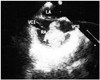
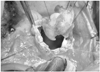
A 24-year-old female patient with a history of primary repair for ASD performed 8 years ago admitted to our clinic for her annual checkups. In her previous primary repair surgery, the defect was closed directly with an atriotomy incision followed by 5/0 polypropylene and 4/0 polypropylene sutures. The patient had attended her ensuing controls every three months for a period of one year, and no pathological findings were identified during this period. There was no history of any drugs being used recently. Physical examination was normal, and routine hematological and biochemical laboratory analysis were within normal levels. Electrocardiogram revealed normal sinus rhythm and right bundle branch block. Transthoracic echocardiography (TTE) revealed normal left ventricular systolic functions, mildly dilated right chambers, minimal mitral regurgitation, mild tricuspid regurgitation, systolic pulmonary artery pressure of 30 mm Hg, and an irregular-shaped mass in the right atrium. On her transesophageal echocardiography (TEE) examination, a 3.7×3.5 cm sized giant pedunculated mobile mass was observed being attached to the septum in the right atrium (Fig. 1). After the procedure, the patient was hospitalized. Chest computed tomography (CT) showed no evidence of pulmonary embolism, and ventilation/perfusion scans indicated no problems. Protein C, protein S, and antithrombin III levels were within the normal range. Venous bilateral Doppler of lower extremity and abdominal ultrasonography results were normal. The patient was scheduled for excision of the mass. Surgery was performed via a median sternotomy by utilizing the cardiopulmonary bypass. Venous drainage was via the superior vena cava and the right femoral vein. The right atrium was being opened, and a large mass filling the entire atrium and obstructing the tricuspid valve was observed. The mass was intimately attached to the free atrial wall, it was irregularly-shaped, and 2.2×4.1 cm in dimension. It had a tanned, gelatinous appearance, and showed multifocal areas of calcification. The entire free wall of the right atrium mass was resected. The patient was weaned off with cardiopulmonary bypass without any difficulty. The mass consistent with thrombus formation originating from the suture line was excised (Fig. 2). Histopathological evaluation was consistent with the organized thrombus.
Case 2
A 42-year-old female, without cardiac complaints, was admitted to our clinic presented with chest discomforts, one month history of exertional dyspnea and persistent dry coughs. She had a history of primary ASD repair 3 years ago, and quitted routine follow-ups. In her previous primary repair surgery, the defect was closed directly with an atriotomy incision followed by 5/0 polypropylene and 4/0 polypropylene sutures. The patient had attended her ensuing controls every two months for a period of one year, and no pathological findings were identified during this period. Biochemical and hematological values were within normal limits. Cardiovascular examination was normal and no pathologic sounds or murmurs were detected. The patient was in normal sinus rhythm and not on any medications. Chest examination elicited few scattered crepitations bilaterally. An image from contrast-enhanced CT pulmonary angiogram demonstrated multiple bilateral pulmonary emboli. TTE revealed normal left ventricular systolic functions, heart chambers within normal size, and a mass in the right atrium. On TEE examination, a 3.2×2.4 cm mobile and irregularly-shaped mass was observed in the right atrium. The patient's bilateral Doppler of lower extremity, abdominal ultrasonography, and hypercoagulability screening panel was negative. Excision of the mass with redo sternotomy was decided for treatment. During the surgery, exploration of the right atrium revealed a 3.2×2.3 cm globular mass with a tanned, gelatinous appearance and multifocal areas of calcification. The mass was attached to the free wall of the right atrium by a 1-cm stalk. This mass originating from the free wall of the right atrium was then excised and its histological examination later revealed it to be an organized thrombus. Both patients had uneventful postoperative courses, and were discharged with warfarin (international normalized ratio: 2-3) combined with acetylsalicylic acid therapy on postoperative seventh and sixth day, respectively. The TTE and TEE showed no thrombus after 6 months of follow-up for both patients.
Discussion
The right atrium contains crista terminalis, eustachian valve and chiari network, and these anatomical structures often cause incorrect interpretations for the evaluation of masses.6) Thrombi, myxomas, and vegetations should be kept in mind for the differential diagnosis of right atrial masses. Fifteen percent of the myxomas derived from the right atrium and are usually linked to the interatrial septum with a broad base and a narrow stalk.7) Myxomas are usually slow-growing, but sometimes may show rapid progressions and may occur after repairs of ASD.8) We offered possible diagnoses among our cases, the right atrial myxoma and thrombus were definitely diagnosed by histopathological examination after surgery.
After the ASD closure, possible complications include pericardial effusion, arrhythmias and thrombus formation. Embolization complications have been reported after ASD closure with transcatheter devices,9) prosthetic patches10) and pericardial patches.11) However, cases of embolization or intracardiac thrombus developed after primary closure of ASD are less common. Right atrial thrombi appearing in the postoperative period is a rare finding and possibly originated from the site of interatrial trauma by suction and devices or suture line. Previously published cases of atrial thrombus detected in the free wall of the right atrium were also detected in the postoperative third month and third year.4)5) In addition, in the report of thrombus on the primary sutured patent foramen ovale site and complicated with systemic embolization by Rodriguez et al.,12) the atrial thrombus was detected six months later after the operation. In the cases which we presented, thrombus were detected at longer periods such as 8 years and 3 years after surgery, and they were treated with surgical interventions.
Right atrial thrombus, which are almost always accompanied by pulmonary embolism, carry a higher mortality rate in the preoperative and perioperative period. Treatment of right chamber thrombi is controversial; medical, surgical procedures and percutaneous interventional techniques can be applied.13) When using thrombolytic agents for the treatment of immobile intra-cardiac thrombus, there is a risk that the dissolution of the peduncule might lead to the migration of the thrombus, which in return may result in embolization.14)15) In addition, one of our patients were complicated with pulmonary embolism. Thrombolytic therapy might have partially disintegrated the thrombus, thus, leaving smaller thrombi. These could have easily migrated into both pulmonary arteries that were nearly completely occluded, therefore, we preferred surgical treatments.
There is no consensus on the duration of anticoagulant therapy after repair of ASD. Anticoagulation treatment for 3 months was previously proposed and implemented.4)16) As seen in our cases, for the detection of thrombus formations over the years after operation, further studies on long-term antiplatelet and anticoagulant therapy are necessary. Right atrial incisions are used in many other intracardiac procedures, such as congenital repair. This complication can thus occur in all surgeries requiring right atrial incision. Therefore, it is important to bear in mind and apply the above mentioned precautions following the completion of these procedures.
The long-term detection of atrial thrombus after operation shows the importance of routine echocardiography. Echocardiographic follow-ups after the procedure are recommended, usually at 24 hours, 1 month, 6 months, 1 year and then, at regular intervals.17)
As a result, thrombosis may occur after closure of ASD in the right atrium. Caution should be exercised in the long-term follow-up period, especially in terms of intra-atrial thrombus.
 XML Download