

 PDF
PDF ePub
ePub Citation
 Print
Print


A 56-year-old man presented to the emergency room with a history of cough and hemoptysis for one week. Two months earlier, he had been diagnosed with dilated cardiomyopathy on the basis of clinical and echocardiographic features. He also reported worsening shortness of breath over the last week. He denied chest pain or fever. His blood pressure was 120/90 mm Hg, heart rate was 108 beats/minute, respiratory rate was 22/minute, body temperature was 97°F, and capillary oxygen saturation was 98% on air. Physical examination revealed bilateral crackles at the bases of both lungs. The on-call physician prescribed antibiotics and diuretics pending further investigation.
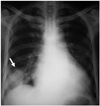
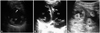
His chest X-ray showed Hampton's hump, a wedge-shaped area of consolidation in the right lower lung field (Fig. 1, arrow), which prompted the suspicion of pulmonary infarction.1) Transthoracic echocardiography showed severe global left ventricular dysfunction and biventricular thrombosis {Fig. 2. LV: left ventricle, RV: right ventricle, thrombi (arrows and asterisks)}. We treated the patient with unfractionated heparin and warfarin. A repeat echocardiogram eight days later indicated that the biventricular thrombi had completely resolved. The patient was discharged when the International Normalized Ratio was 3.2 on oral anticoagulation.
The diagnosis of pulmonary embolism is particularly challenging in low income settings due to expensive investigations, such as computed tomography, which are not routinely available.2) In this case, the rare finding of biventricular thrombosis on echocardiography together with Hampton's hump on chest X-ray was helpful in establishing the diagnosis of pulmonary embolism and infarction.3)
 XML Download