

 PDF
PDF ePub
ePub Citation
 Print
Print


Introduction
Multivessel myocardial infarction (MI) is a rare case. Most cases of multivessel MI are simultaneous events and consecutive events of MI usually involving the same vessel. A case with consecutive events of acute MI in different vessels is rare. In addition, patients of consecutive events of acute MI have multiple risk factors or specific underlying diseases. There have been no reports on 3 consecutive events of acute MI in each 3 vessels during a long-term interval. Herein, we report the first case with 3 consecutive events of acute MI in 3 different vessels during a long-term interval.
Case
On September 25, 2012, a 51-year-old man visited the emergency room for acute retrosternal squeezing chest pains for the past twelve hours. He had no history of diabetes mellitus, hyperlipidemia, and nor a familial history of coronary artery disease. However, he was a current smoker (30 pack years). He had hypertension and history of two consecutive acute MI.
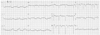
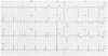
The first acute MI was on February 21, 2000. The electrocardiogram (ECG) showed ST-segment elevations in the inferior leads (II and aVF) and the anterior (V 2-4) leads (Fig. 1). The peak levels of creatine kinase-MB (CK-MB) and troponin T were 31.71 and 2.51 ng/mL. The coronary angiography was performed. The coronary angiography showed a total occlusion with thrombus and Thrombolysis in Myocardial Infarction (TIMI) 0 distal flow in the proximal portion of the left anterior descending from the coronary artery (LAD), which was successfully treated with a bare metal stent (3.0×15 mm, NIR Royal® stent, Boston Scientific Corporation, Natick, MA, USA). In addition, he had taken aspirin 100 mg, atenolol 25 mg, captopril 37.5 mg and simvastatin 10 mg once daily and cilostazol 100 mg twice daily, on a regular basis after the first MI event.
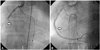
The second acute MI was on December 30, 2007. The ECG showed ST-segment elevation in the inferior (II, III, and aVF) leads (Fig. 2). The peak levels of CK-MB and troponin T were 164.7 and 4.01 ng/mL. Emergent coronary angiography was performed. The coronary angiography showed total occlusion with thrombus, with TIMI 0 distal flow in the mid portion of the right coronary artery (RCA), which was successfully treated with a paclitaxel eluting stent (4.0×24 mm, TAXUS®, Boston Scientific Corporation, Natick, MA, USA) (Fig. 3-1), and a 95% restenosis in a stent with thrombus, and TIMI 0 distal flow in the proximal portion of the LAD, which was successfully treated with a zotarolimus eluting stent (2.75×30 mm, ENDEAVOR®, Medtronic Inc., Minneapolis, MN, USA) and overlapping a sirolimus eluting stent (3.0×23 mm, CYPHER®, Cordis Corporation, Warren, NJ, USA) (Fig. 3-2). There were no other stents for appropriate lesion lengths and sizes, except ENDEAVOR and CYPHER stents at that time.
He had taken aspirin 100 mg, clopidogrel 75 mg, ramipril 2.5 mg and simvastatin 10 mg once daily and molsidomine 2 mg twice daily, on a regular basis after a second MI event.
At the third acute MI, physical examination showed that his blood pressure was 90/60 mm Hg, the heart rate was 64/min, and respiratory rate was 20/min. He had a regular heart beat and clear heart sounds without murmurs.
Electrocardiogram showed Q-wave and ST elevation in the V 1-5 and T-inversion in V 4-6 (Fig. 4). The peak levels of CK-MB and troponin T were 17.84 and 0.741 ng/mL.
Echocardiographic evaluation revealed aneurysmal changes at the apicoseptal wall in the left ventricle, and ischemic insult in the LAD and the RCA territory. An ejection fraction of 40% was observed.
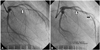
Emergent coronary angiography showed a total occlusion with thrombus and TIMI 0 distal flow in the proximal portion of the left circumflex coronary artery and collateral flow from RCA, which was successfully treated with a zotarolimus eluting stent (3.5×22 mm, RESOLUTE INTEGRITY®, Medtronic Inc., Minneapolis, MN, USA) (Fig. 5). After primary percutaneous interventions, the patient became stable without chest pains and was discharged on the fourth hospital day.
Hematological analyses were performed and showed the following results: D dimer 0.38 ug/mL (<0.5 ug/mL), antithrombin III 107% (80-120%), protein C activity 131% (70-130), and protein S activity 90% (70-130). In addition, immunological analyses were performed and indicated the following results: lupus anticoagulant titer 1.01 (0.8-1.2), anti-beta2-glycoprotein1 immunoglobulin G/M titer negative/negative, anti-cardiolipin antibody immunoglobulin G/M titer negative/negative and homocystein 12.33 umol/L (5.08-15.4 umol/L). Further, the platelet drug response assay was also performed and displayed the following results [The results of the assay are reported as absolute P2Y12-Reaction-Units (PRU), as well as percent inhibitions (% inhibition), the latter calculated as 100-(TEST/BASE×100) or {(BASE-TEST)/BASE}×100]: PRU 215 (0-240), % inhibition 31% (20-80).1) He was discharged after uneventful recovery with triple anti-platelet therapy using aspirin 100 mg and clopidogrel 75 mg once daily and cilostazol 100 mg twice daily. He had also been treated with ramipril 2.5 mg, bisoprolol 2.5 mg and rosuvastatin 10 mg once daily and molsidomine 2 mg twice daily. The patient has been followed-up at the outpatient department without further symptoms.
Discussion
Coronary artery thrombosis is the pathogenic mechanisms of MI. The formation of an intraluminal clot is a result from the loss of integrity of a protective covering over an atherosclerotic plaque. The loss of integrity from the protective covering induces plaque ruptures or erosions. This disruption allows blood to come in contact with the highly thrombogenic contents of the necrotic core of the plaque and luminal thrombosis to occur.2)3)
There are many risk factors of acute MI, such as cigarettes smoking, hypertension, dyslipidemia, diabetes mellitus, family history of coronary heart diseases, old age, obesity, physical inactivity, and atherogenic diets. Other rare risk factors of acute MI are hyperhomocysteinemia, dysregulated coagulation and fibrinolysis. However, he had no conventional risk factor, except for smoking. There was also no abnormal values for homocystein, coagulation factors, and inflammatory factor.
The second MI event was de novo lesion and stent thrombosis. Several factors have been associated with stent thrombosis, including older age, black race, diabetes mellitus, bifurcation lesion, instent restenosis lesion, and other procedure-related factors, such as stent malposition, greater stent length, post-procedure acute renal failure, non-compliance to anti-platelet agent and anti-platelet resistance.4) Clopidogrel resistance has been defined as incomplete blockade of the platelet membrane P2Y12 receptor, as measured by various laboratory tests specifically to clopidogrel's mechanism of platelet inhibition according to a patient compliant with clopidogrel therapy.5-7) We checked the patient's platelet drug response assay, but there was no abnormal values. Furthermore, we also checked the patient's compliance of medication; he had good compliances.
We reviewed other cases of acute MI, including consecutive events or simultaneous events, as well as for same vessels or different vessels. Some cases were reported simultaneously with multi-vessels coronary artery thrombosis.8-10) Several possible underlying conditions for coronary thromboses have been suggested, such as cocaine use, hypercoagulable state and essential thrombocytosis. In addition, most patients had multiple risk factors for coronary artery diseases. Acute multiple coronary thromboses may be associated with a systemic prothrombotic condition. Another mechanism is that the first event, causing impairments of other vessel flows, can lead to acute secondary thrombosis. The other mechanism is due to aortic or mitral valve endocarditis. Some cases on consecutive events of acute MI were also reported. The reasons of consecutive acute MI are clopidogrel resistance,11) anatomical abnormality (papillary fibroelastoma12)), hematologic abnormality (antithrombin III deficiency13)), or other specific underlying diseases. One case was reported with two consecutive events of acute MI in two different vessels during a four-year interval. The patient in the above mentioned case has the Budd-Chiari syndrome and Behcet's disease.14)
There was no reported case for three consecutive events of acute MI in three different vessels during a long-term interval. The evaluation of the causes for consecutive MI attack is important in order to prevent further coronary vascular events which could happen in such manner. However, he did not have risk factors, such as coagulation abnormality, clopidogrel resistance, patient's compliance and vessel abnormality, except for smoking. He has already stopped smoking, thus, there was no correctable factor. So we treated him with triple antiplatelet therapy and planned short term interval follow-ups.

 XML Download