

 PDF
PDF ePub
ePub Citation
 Print
Print


Introduction
An aberrant right subclavian artery (ARSA), also called "arteria lusoria", was first described by Hunauld from autopsy studies in 1735.1) It is the most common vascular abnormality of the aortic arch, occurring in 0.7-2.0% of the population.2) This anomaly is a result of abnormal regression of the fourth aortic arch and persistent patency of the eighth right dorsal aortic segment.3) Generally, ARSA remains asymptomatic. However, ARSA has been associated with development of an aneurysm in approximately 3-8% of these patients, accompanied by dysphagia and upper airway symptoms due to the compression of trachea and esophagus. Eventually, it may be fatal due to its marked propensity to rupture.1) In the past, surgical repair of the aorta has been the mainstream of treatment for the aortic arch diseases. However, with advance of endovascular skills and devices, a hybrid procedure combining less-invasive surgery with endovascular therapy using stent grafts to reduce the risk of procedure-related morbidity and mortality is gaining attention.
In this case report, we describe an elderly patient with a thoracic aortic aneurysm and ARSA who was successfully treated with a hybrid procedure of surgical replacement of ascending artery, bilateral carotid-to-subclavian artery bypass and implantation of stent graft in the aortic arch and descending aorta.
Case
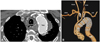
An 84-year-old man presented with chest pain on left side. He was a smoker of 60 years' standing and had been previously diagnosed with hypercholesterolemia. However, he had no past history of hypertension or diabetes. A chest X-ray showed markedly dilated aortic notch. Computed tomography (CT) aortography revealed an ARSA originating from the distal aortic arch, distal to the left subclavian artery and crossing the mediastinum between esophagus and trachea, and a thoracic aortic aneurysm with a diameter of 67 mm and a thrombus extending from the left subclavian artery origin to the proximal descending aorta involving the right subclavian artery origin (Fig. 1). The ascending aorta was also dilated with a maximal diameter of 40 mm and showed partial calcification on the anterior aorta wall. Electrocardiogram was nonspecific. Echocardiography showed normal sized cardiac chambers with left ventricle ejection fraction of 74%. Thoracic aortography confirmed the CT findings. Coronary angiography demonstrated 50% narrowing of the proximal left anterior descending artery. CT brain also revealed that the circle of Willis and both vertebral arteries were patent. Results of laboratory tests showed elevated total serum cholesterol (237 mg/dL) and low density lipoprotein cholesterol levels (164 mg/dL).
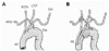
We decided to perform a staged hybrid procedure combining surgical replacement of the ascending aorta and bilateral carotid-tosubclavian artery bypass with implantation of a stent graft in aortic arch and descending aorta. In the first stage of the procedure, the ascending aorta was replaced by a graft. Under general anesthesia, median sternotomy was performed. Cardiopulmonary bypass was performed after cannulation of the right axillary artery and the right atrium. The ascending aorta was clamped and the heart was arrested with cold antegrade cardioplegia infusion. After selective antegrade cerebral perfusion was started, the ascending aorta was replaced using 4-branched hemashield graft (28, 10, 8, 8, 10 mm, Boston Scientific, Wayne, NJ, USA). Two 10 mm graft branches were ligated. The other two 8 mm graft branches were anastomosed with each of the common carotid arteries. The cardiopulmonary bypass, aortic across clamp, and circulatory arrest time was 162, 87, and 15 minutes, respectively. For securing a sufficient landing zone for stent graft implantation, the distal end of the hemashield graft was inserted into distal aortic arch as an elephant trunk. In our patient, the elephant trunk is attached to the proximal aorta arch after debranching and bypassing both common carotid arteries from the hemashield graft of ascending aorta in order to exclude dilated aorta arch distal to the origins of the left and right subclavian arteries (Fig. 2).
The second procedure was performed 14 days after the first procedure. Under general anesthesia, a left neck skin incision was made. Left common carotid and left subclavian arteries were exposed. A bypass surgery connecting common carotid artery and subclavian artery was performed using an 8 mm hemashield graft. A bypass surgery connecting the right common carotid artery with the subclavian artery was performed 6 days later as a third stage procedure because of fragile patient's condition after the surgery. After the closure of the incision site, the patient was placed under a fluoroscope. Both common femoral arteries are punctured. A 5 Fr marker pigtail catheter was inserted through the left femoral artery for the angiographic guidance. The right femoral artery was prepared for the later closure using two Perclose ProGlide suture closure devices (Abbott Vascular, Redwood City, CA, USA). Amplatz Vascular Plugs II (14 mm in diameter, AGA Medical Corporation, Golden Valley, MN, USA) were implanted into the right and the left subclavian artery using a 7 Fr shuttle sheath in order to prevent retrograde endoleaks into aortic aneurysm. A 0.035 inch extra stiff wire (Lunderquist, Cook Inc., Bloomington, IN, USA) was inserted through the right femoral artery into the ascending aorta. A 34×202 mm thoracic stent graft (Zenith TX2, Cook, Bloomington, IN, USA) was inserted over the wire and deployed within the hemashield graft implanted in the aortic arch. Stent grafts of appropriate size were not available from one vendor at that time. Therefore, an additional 40×100 mm stent graft (SEAL, S & G, Seoul, Korea) was implanted distal to the first stent graft in an overlap manner. A 40 mm balloon (Coda, Cook Inc., Bloomington, IN, USA) was used to appose the stent graft the aorta wall under rapid pacing. An immediate post-implant aortography showed no significant endoleak (Fig. 3). The right femoral artery access site was closed by tightening the knots prepared by Preclose technique without complication. At a CT taken before discharge, the aneurysm was completely excluded without significant endoleak. The patient was discharged in good status 5 days after the stent graft procedure and in the subsequent 2 years has remained well with no complications (Fig. 4).
Discussion
Normally, the fourth right aortic arch forms the right subclavian artery. However, with regression of the fourth right aortic arch and the right dorsal aorta, the ARSA develops as the fourth branch of the aortic arch distal to the left subclavian artery in this anomaly due to the persistence of the seventh inter-segmental artery.3) In 80% of the individuals with an ARSA, it crosses between the esophagus and the vertebral column.4) However, it runs between the esophagus and the trachea in 15% of the cases. In 5%, it passes anterior to both the trachea and esophagus.5) Sixty percent of patients with ARSA have Kommerell's diverticulum, an aortic diverticulum at the site of origin of the aortic arch.4) Based on previous reports, patients presenting with an ARSA aneurysm require treatment if the aneurysmal sac is larger than 3 cm.6) The classic treatment method for ARSA involves surgical exclusion of the ARSA and revascularization of the right arm, usually requiring thoracotomy, extracorporeal circulation, selective brain perfusion, and deep hypothermia.7) However, surgical repair of ARSA is challenging due to significant risk of neurologic complications and high mortality rates ranging from 9 to 50%.8) Generally, endovascular repair of the aortic arch remains a technical challenge. Several approaches such as implantation of fenestrated or branched stent grafts or using the chimney graft technique for preserving blood flow to aortic arch branches are reported to be feasible. Nevertheless, these techniques are not usually applicable to ARSA aneurysms. Therefore, a hybrid rocedure of less-invasive surgery and endovascular therapy to avoid severe complications and mortality may be an attractive alternative therapy especially for patients at high surgical risk. Generally, hybrid treatment consists of endovascular exclusion of the ARSA using a stent graft and revascularization of subclavian arteries in staged procedures. Since Lacroix demonstrated the first case of successful hybrid treatment of an aneurysmal ARSA, several authors have reported cases treated using this technique.7)9-11) In the present case, surgical replacement of the ascending aorta was required due to its aneurysmal change. Therefore, aortic arch branches were surgically debranched and connected with ascending aorta using branched grafts to ensure sufficient landing zone for stent graft implantation in the aortic arch. Because both subclavian arteries were about to be covered by stent grafts, we revascularized bilateral subclavian arteries by bypassing the subclavian artery to the common carotid artery on each side prior to the implantation of stent graft. In general, the revascularization of the subclavian arteries are not always required if the circle of Willis is intact and the ipsilateral vertebral artery is not dominant in supplying blood flow to the other side. However, revascularization of the subclavian artery is recommended whenever possible because of potential risk of cerebral, upper extremity, and spinal cord ischemia.
In conclusion, the present case demonstrated that a less invasive hybrid treatment of endovascular therapy and less extensive surgery can be performed successfully for the treatment of ARSA with aneurysmal change. We believe this is an effective alternative treatment for patients at high surgical risk.

 XML Download