

 PDF
PDF ePub
ePub Citation
 Print
Print


Introduction
The drug-eluting stent (DES) has changed our approach to the management of acute myocardial infarction (AMI) over the past decade. The first commercial DES was a significant innovation because it reduced the numerous drawbacks of the bare-metal stent (BMS), including high in-stent restenosis (ISR) rates with neointimal hyperplasia (NIH) as an underlying mechanism.1-5) Drugs coating the stents were categorized as cytotoxic agents, such as paclitaxel, a chemotherapeutic agent in the treatment of breast or gastric cancer, and sirolimus, an anti-inflammatory agent in kidney transplantation, that were used in first-generation DESs. Commonly used second-generation drugs in stents are zotarolimus, an immunosup pressant and semisynthetic derivative of rapamycin, everolimus, a derivative of sirolimus that works as an inhibitor of the mammalian target of rapamycin (mTOR), and biolimus, an equipotent semi-synthetic sirolimus analog with a biodegradable polymer, polylactic acid, with ten times the lipophilicity of sirolimus. All three types of DES have improved clinical outcomes after percutaneous coronary intervention (PCI) by reducing ISR, stent-thrombosis, and the duration of anti-platelet therapy.6-8)
The Heart Research Center of the Chonnam National University Hospital has investigated the safety and efficacy of various drug-coated stents in both human and animal studies (Fig. 1). Some demonstrated usefulness in avoiding ISR by preventing neo-intimal regrowth. In the present review, we cover imported DESs and the new DESs that have been developed in our center, and provide perspectives on DES development.
Searching for Optimal Coating Materials
Conventional coating materials from western countries
The first generation: paclitaxel and sirolimus
Paclitaxel is a mitotic inhibitor used to treat patients with lung, ovarian, breast, and head and neck cancer, as well as advanced forms of Kaposi's sarcoma. Sirolimus, also known as rapamycin, is an immunosuppressive drug used to prevent organ rejection after transplantation. It is particularly useful in kidney transplants.9)
The second generation: everolimus and zotarolimus
Everolimus is the 40-O-2-hydroxyethyl derivative of sirolimus and works similar to sirolimus as an inhibitor of mTOR. It is currently used as an immunosuppressant to prevent organ rejection after transplantation and to treat renal cell carcinoma and other tumors. Zotarolimus is an immunosuppressant and a semi-synthetic derivative of rapamycin. It was designed for use in stents with phosphorylcholine as a carrier.10)
Experimental coating materials at Chonnam National University Hospital
Heparin
Heparin, an anticoagulant, can interact with antithrombin III to prevent thrombus formation and activation.11) Heparinization of surfaces has proven to be a successful strategy in preventing thrombus formation and improving the compatibility of blood-contacting biomaterials.12-14) Heparin has anticoagulant properties and inhibits smooth-muscle cell (SMC) proliferation. Local drug delivery with heparin-coated stents can deliver a high concentration of heparin to lesion sites without systemic bleeding complications.
Abciximab
At the beginning of the 21st century, investigators in our group examined the effectiveness of abciximab, not only as an adjunctive agent during PCI, but also as an eluting material in the manufacture of coronary stents. Our group conducted many investigations on human as well as animal subjects with the Reopro® (abciximab)-coated stent. The results will be discussed elsewhere in this review. Abciximab, a potent antiplatelet agent that blocks the ultimate pathways to platelet aggregation, improves the outcomes of high-risk PCI and decreases the incidence of major adverse cardiac events.15-18) In contrast to other types of platelet glycoprotein IIb/IIIa receptor blockers, it also binds to Mac-1 (CD11b/18) on vascular endothelial cells and macrophages, thereby inhibiting inflammatory responses and SMC proliferation after vascular injury.19-24)
Ramiprilat
The renin-angiotensin-aldosterone system has been implicated in the pathogenesis of NIH. Angiotensin II has been found to be associated with the migration and proliferation of vascular SMCs in restenotic lesions. Moreover, the administration of angiotensin converting enzyme inhibitors (ACEIs) has been reported as reversing impaired endothelium-dependent responses to acetylcholine in patients with heart failure25)26) and hypertension, as well as improving coronary endothelial function after treatment with an ACEI in patients with coronary artery disease (CAD).27)28)
Alpha-lipoic acid
Alpha-lipoic acid (α-LA) is a potent antioxidant and acts as a cofactor of key mitochondrial enzymes, such as pyruvate dehydrogenase and α-ketoglutarate dehydrogenase.29) α-LA is endogenous and improves diabetes-induced endothelial dysfunction, probably due to its antioxidant effects and direct free-radical scavenging properties.30) Moreover, α-LA inhibits the inflammatory pathway and prevents NIH after carotid artery stenting.31)32)
Carvedilol
Carvedilol is a neurohumoral antagonist with multiple actions. It was originally discovered as a β-adrenoceptor antagonist, but subsequent research revealed that it also possesses potent antioxidant and free-radical scavenger properties. In addition, carvedilol inhibits vascular smooth muscle cell (VSMC) proliferation induced by a broad group of mitogens, such as platelet-derived growth factor, fibroblast growth factor, endothelin-1, serum, and thrombin, and was found to suppress 84% NIH in a rat carotid injury model.33)34) Recent studies have shown that the antimitogenic mechanism of the action of carvedilol on VSMCs involves the inhibition of mitogen-activated protein kinase activity and the regulation of cell cycle progression. Carvedilol is highly lipophilic, which promotes rapid cellular uptake,35)36) and a stent containing carvedilol allows a high local concentration of the drug to reach the vessel walls with which it comes into contact.
Probucol
Probucol was developed and sold as a lipid-lowering agent and is also used as an antioxidant,37) reducing restenosis after percutaneous transluminal coronary angioplasty. It has, furthermore, recently been identified as a vascular protectant.38-41) A multicenter study recently reported that post-stenting restenosis was reduced after probucol use via a mechanism that increases the diameter of the vessel lumen.42)43) The combination of probucol and candesartan has potential antioxidant and vascular protectant effects.44)
Fucoidan
Fucoidan is a sulfated polysaccharide extracted from brown seaweed that reduces rat SMC proliferation in vitro in a more intensive manner than heparin.45) Fucoidan inhibits SMC proliferation by reducing mitogen-activated protein kinase activity. Investigators at our center recently succeeded in producing and characterizing new homogeneous fractions of low-molecular-weight fucoidan with low anticoagulant activity. They aimed to establish the optimal conditions for the fucoidan coating on a BMS, verifying both the ability of fucoidan to inhibit vascular SMC proliferation in vitro and the inhibitory effect of a fucoidan-coated stent on ISR in a porcine model.46)
Development of a coating method
Dopamine-mediated heparin-coated stent
In recent decades, typical approaches to surface heparinization, such as layer-by-layer self-assembly and covalent immobilization, have been reported in medical literature.47-49) Layer-by-layer self-assembly has a common limitation involving the clinical translation of materials functionalized with biomolecules. That is, these coatings fail to withstand long-term exposure in vivo because of a lack of robustness. Inspired by the composition of adhesive proteins in mussels, Bae et al. used dopamine to form thin and surface-adherent films on a stent surface.50) Dopamine performs well as a binding agent for coating inorganic surfaces, including stent materials, and others have reported improved hydrophilicity and a substantial reduction of protein adsorption by dopamine.50)
Titanium dioxide-coating method
We had discovered that the thin film of titanium dioxide (TiO2) was deposited onto a BMS through the plasma-enhanced chemical vapor deposition (PECVD) process. We investigated the potential of TiO2 as a drug-combining matrix. When deposited at a discharge power of 5 W, the film showed a highly smooth surface with a roughness of 9.4 nm, mechanical stability with good adhesion, and good blood compatibility. The film was surface modified with water plasma to introduce hydroxyl groups on the TiO2 surface. Drugs were then able to be chemically grafted onto the modified surface through the formation of ester bonds between hydroxyl groups on the modified TiO2 film, and carboxyl groups in the drugs. When heparin, α-LA, and abciximab were grafted onto the TiO2-coated and surface modified stents, the mean engrafted amount was measured at 106.1 mg for α-LA, 32.5 mg for abciximab, and 53.9 mg for heparin. In the in vitro drug release test, heparin and abciximab were released continuously for four weeks, but α-LA exhibited a burst release within six days.53)
Nitrogen oxide-doped titanium dioxide-coated stent
Titanium dioxide or nitrogen-doped titanium oxide (N-TiO2) coatings, inter alia, have been studied intensively as biomedical implants for contact with blood in organs, such as heart valves, or in vascular stents, as they inhibit platelet aggregation and fibrin growth. TiO2 film could be applied as a drug-binding matrix as an alternative to organic polymers in DESs. Drugs can be directly grafted onto a TiO2 film surface through the chemical bond formation between functional groups of the drug and -OH groups of plasma modified TiO2 film. N-TiO2 film is especially attractive as a coating material among blood-contacting materials, because of its enhanced blood compatibility.54-56)
Such films are not yet being used as coating for drug binding matrices in DES. Therefore, we discussed the use of N-TiO2 film coatings for DES as a drug-combining matrix, by investigating the properties of coating films prepared by PECVD and by optimizing the surface modification of the N-TiO2 film coatings. N-TiO2 films were promising alternatives to organic polymers in the preparation of DESs, because grafted heparin and abciximab can be released continuously for three weeks from drug-grafted N-TiO2 stents in in vitro drug release testing.57)
Dual-coated stent (abciximab and alpha-lipoic acid)
Abciximab (Reopro®) is a potent anti-platelet agent, α-LA (a potent antioxidant), and acts as a co-factor of key mitochondrial enzymes, such as pyruvate dehydrogenase and α-ketoglutarate dehydrogenase.58) It improves endothelial function and prevents atherosclerosis-related disease.29) We compared the effect of a stent coated with abciximab and α-LA with that of BMS in a porcine coronary overstretch restenosis model. The findings are provided elsewhere in the present review.
Aptamer stent: endothelial progenitor cell capture stent
Aptamers are oligonucleic acid or peptide molecules that bind to a specific target molecule that are usually selected from a large random sequence pool. However, natural aptamers also exist in riboswitches. Aptamers can be used for both basic research and clinical purposes as macromolecular drugs.
Aptamers can be combined with ribozymes to self-cleave in the presence of their target molecules. These compound molecules have additional research, industrial, and clinical applications. The aptamer stent was specifically designed to promote arterial healing using a coating of oligonucleotides, functioning as endothelial progenitor cell (EPC) attracting messengers. As a result, the aptamer stent captures and sequesters circulating EPCs to the luminal stent surface and theoretically initiates re-endothelialization.59)
Results of clinical trials of conventional drug-eluting stents
Animal studies and clinical trials of Chonnam National University Hospital stents
Heparin-coated stents
Heparin-coated stents were compared with control BMS stents in a porcine coronary stent restenosis model with stent overdilation injury (stent : artery=1.3 : 1.0). The heparin-coated stent was effective in the prevention of late coronary stent restenosis in a porcine model, indicating a possible relationship with the inhibition of neointimal cell proliferation.5) In 2001, Shin et al. showed that heparin-coated stents were safe in the early setting of AMI. No additional heparin infusion after stenting was necessary, potentially reducing the risk of bleeding complications.46) In 2004, Madduri et al. reported that the use of a heparin-coated stent resulted in favorable procedural and six-month outcomes, no incidence of stent thrombosis, and overall positive cardiac prognosis at the six-month follow-up.29) However, Mehran et al. conducted an internet-based registry examining the efficacy of a heparin-coated stent, and found no significant difference in cardiac death, myocardial infarction, target-lesion revascularization, or stent thrombosis between a heparin-coated stent versus BMS.53)
Abciximab-coated stents
Abciximab-coated stents have been shown to be safe and effective in the prevention of coronary restenosis in humans, as well as in a porcine model.32)71) A study of 85 patients undergoing follow-up coronary angiography and an intravascular ultrasound (IVUS) showed only a trend toward a decreased restenosis rate. However, late luminal loss was lower in the abciximab-coated stent group.5) In a study of 150 patients, the restenosis and target-vessel restenosis rates of an abciximab-coated stent were lower than those with a conventional BMS.72) Later, in a study of 96 patients, follow-up IVUS demonstrated an improved intrastent luminal area and intrastent NIH. The ISR rate was lower in the abciximab-coated stent group than in the conventional BMS group.73) In an examination of 138 stents (69 abciximab-coated stents vs. 69 BMS), the restenosis rate and late loss rate were both lower in the abciximab-coated stent group than in the BMS group. At the follow-up IVUS, the intrastent luminal area was significantly larger, and the intrastent NIH area significantly smaller, in the abciximab-coated stent group than in the BMS group.74) In addition, abciximab-coated stents demonstrated anti-inflammatory effects in a study using a porcine coronary overstretch restenosis model in a histopathological analysis performed 28 days after stenting with abciximab-coated stents versus SES or PES.75) However, Kim et al.76) conducted a prospective randomized study comparing the effects of the abciximab-coated stent in 95 patients with BMS with 93 control patients after a two-year follow-up. The six-month IVUS analysis showed that the area of NIH was significantly smaller in the abciximab-coated stent group than in the control group. Nevertheless, at the two-year clinical follow-up, there were no statistically significant differences between the groups in terms of clinical outcomes.
Ramiprilat-coated stent
We examined the anti-proliferative and anti-inflammatory effects of ramiprilat-coated stents in a porcine coronary overstretch restenosis model and observed no significant differences in the injury and inflammation scores between the two groups. Within the neointima, most inflammatory cells were lymphohistiocytes. Although the ramiprilat-coated stent did not demonstrate significant inhibitory effects on NIH, this stent showed positive effects on inflammatory reaction and arterial healing, similar to the control stent in a porcine coronary restenosis model.77)
Alpha-lipoic acid-coated stent
Lim et al.,71) in our center, found that α-LA feeding and α-LA-coated stents prevent ISR by inhibiting NIH in a porcine coronary artery restenosis model, possibly by inhibiting the activation of the NF-κb pathway and the proliferation of porcine VSMCs.
Carvedilol-loaded stents and probucol-coated stents
Kim et al. utilized BiodivYsio® phosphorylcholine-coated stents, dip-coated with carvedilol (5 mg/mL) or probucol (50 mg/mL) by immersion, in respective methanol solutions. Twenty-four stents (carvedilol=8, probucol=8, and control=8) were placed in twelve pigs. Histopathological analysis was performed four weeks later. A histomorphometry of the carvedilol-coated stent group compared with the control group showed that the neointimal area decreased by 42% and the lumen area increased by 20%, resulting in a 43% reduction of the % stenosis area. In the probucol-coated stent group, the lumen area, neointimal area, and % stenosis area did not differ significantly from those of the control group. The carvedilol-coated stent, but not the probucol-coated stent, inhibited NIH in a porcine stent restenosis model.29)
Fucoidan stent
Fucoidan-coated stents (FCSs, fucoidan 12.5 mg/mL), PESs, and BMSs were implanted in the proximal left anterior descending artery, proximal left circumflex artery, and right coronary artery in a randomized manner in six pigs (three stents placed in three coronary arteries per pig). Histopathological analysis was performed on the 28th day of stenting. No polymer cracks or blotched areas were observed in the dual coating. Proliferation of SMC was inhibited in the fucoidan-treated group (12 µg/mL) compared to the control group. There were no significant differences in the internal elastic lamina, lumen area, neointimal area, or % stenosis area among the three stent groups. On the BMS surface, dual coating was appropriate using fucoidan. Despite the fucoidan inhibited SMC proliferation in vitro, the FCS showed no significant differences in the histopathological analysis performed.46)78)
Abciximab and alpha-lipoic acid (a dual-coated stent)
Lim et al. examined the anti-proliferative and anti-inflammatory effects of a stent coated with abciximab and α-LA in a porcine coronary overstretch restenosis model. A total of ten pigs were randomized into two groups (ten pigs, ten coronaries in each group), in which the coronary arteries were stented with a dual-coated stent and a BMS (control) after randomization. There was no significant difference in the injury score between the two groups. In the neointima, the lymphohistiocyte count was significantly lower in the dual-coated stent group than in the control stent group. There was no significant difference in the fibrin score between the two groups. The neointima area was not significantly different between the two groups. Although the dual-coated stent with abciximab and α-LA showed no significant difference in terms of the inhibition of NIH when compared with the BMS, it was associated with a reduced inflammatory reaction when compared with the control stent in a porcine coronary restenosis model.79)
Endothelial progenitor cell capture stent (an aptamer stent)
We evaluated the effects of the aptamer-coated stent utilizing six pigs and three different stents: cobalt-chromium (CC) BMS, paclitaxel eluting Taxus PES (Boston Scientific, Natick, MA, USA), and aminoparylene and aptamer (AA)-coated stents, implanted in the three epicardial coronary arteries in a random manner at 8 to 10 atm. Stent length and diameter were 18 mm and 3 mm, respectively. After one month, the animals underwent a follow-up coronary angiography with acetylcholine (10 mL, 3 µg/mL) stimulation tests and were then euthanized for histological analysis. There was only a trend toward a decreased minimal lumen diameter in all stents. Neointimal thickness was lower after the use of Taxus PES. The AA-coated stent group showed a tendency toward a lower incidence of stent thrombosis and endothelial dysfunction compared to the Taxus PES group. However, the inhibition of NIH did not differ between the two groups.59)
Future direction: promising stents
Biodegradable polymer stents
Drug-eluting stents made with biodegradable polymers have been shown to improve long-term safety and efficacy compared to SES. DESs with biodegradable polymers demonstrated improved safety, compared to BMS in patients with AMI,80) and showed outcomes similar to those with EES in patients with CAD at 1 year.81)
Polymer-free drug-eluting stent
Under the hypothesis that the polymer stent coatings reduce late events, polymer-free DES was developed. BioFreedom stents by Biosensors and drug-filled stents by Medtronics do not require polymer coatings on stents. Rather, the drug-filled stent has numerous elution holes through which the drug exits, thereby controlling drug elution through diffusion physics. Potential advantages are 1) avoidance of long-term late adverse effects that might be attributable to the polymer, 2) improved surface integrity since there is no polymer to be sheared or peeled away from the stent struts, and 3) the need for a shorter duration of dual antiplatelet therapy.
A promising drug-eluting stent from Chonnam National University Hospital
Gene delivery stents: Akt1 siRNA-embedded stents
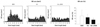
Akt1 siRNA was delivered to the cells with enhanced proliferation to suppress the Akt1 protein responsible for cellular proliferation at the mRNA level.84) The inhibition of Akt1 decreases the expression of caspase-8 in endothelial cells. Suppression of downstream Akt1 signaling proteins such as Fas ligand results in the stimulation of caspase activity and apoptosis in VSMCs. The specific inhibition of Akt1 protein expression has been reported to retard cell growth.85-89) To evaluate the usefulness of a gene delivery stent, rabbit iliac artery stenting was performed at our center. VSMC proliferation over the stent was suppressed by the delivery of Akt1 siRNA complexed with a biodegradable disulfide cross-linked polyethyleneimine (ssPEI) polymer on a hyaluronic acid-coated stent surface. The study results confirmed that the rate of restenosis was slower when stents were coated with ssPEI complexed with Akt1 siRNA (Fig. 2).84)
Drug and gene delivery stents: Abciximab-Kruppel-like Factor 4-plasmid dual-delivery TiO2-coated coronary stents
Recently, we developed a novel drug and gene dual-delivery system for coronary stents using TiO2, with nonpolymer thin film with high blood compatibility used for drug delivery, and a denatured polymer coating for local gene delivery. Our results suggested that this novel system could be useful for the dual delivery of a drug and gene in coronary stents and other biomedical devices. To analyze the inhibitory effect on ISR, both TiO2-abciximab-Kruppel-like factor 4-plasmid-cobalt chromium (TAK-CC) and TiO2-only cobalt chromium (T-CC) stents were used. On histomorphometric analysis after four weeks of implantation in a porcine coronary artery, the TAK-CC group had decreased ISR compared with the T-CC group (TAK-CC group: 26.38±10.79% vs. the T-CC group: 48.59±20.17%, n=6 in each, p<0.05). Neither the TAK-CC nor T-CC stent group showed fibrin deposition by Carstairs' stain, and both groups showed accelerated re-endothelialization, as indicated by detectable anti-CD31-positive stained cells compared with a commercially available PES.90)
Conclusion
The conventional requirements for developing a new DES have been considered to include 1) the inhibitory efficacy of eluting agent against the development of NIH, 2) the stability of the coating agent on the metal stent strut, and 3) cost-effectiveness. However, currently, we must consider DESs with bioabsorbable polymers, such as the ssPEI polymer, in our gene-delivery stent, or polylactic acid, in the biolimus-A9-eluting stent; a polymer-free DES, such as the drug-filled stent by Medtronic; or the development of recent bioresorbable vascular scaffolds. We established a stent factory several years ago and are performing research on the Akt1 siRNA-embedded stent, which is based on biodegradable ssPEI polymer and genetic modification, as well as a drug and gene delivery stent, the TAK-CC. Notwithstanding these efforts, the development of a highly efficient domestic DES may require some time. With cooperation between medicine and mechanics, as well as industry and educational organizations, the fulfillment of these requirements will lead to a commercial domestic DES, benefiting numerous cardiovascular patients in Korea. These benefits would include cost-effective coronary stents due to lower taxes and expenses in the manufacture of coronary stents with safety and efficacy comparable to imported stents. In addition, we are confident that, in the near future, the safety and efficacy outcomes of domestic DESs will be better than those of imported stents.
 XML Download