

 PDF
PDF ePub
ePub Citation
 Print
Print


Introduction
The aorta-right atrial tunnel was first described in 1980 by Coto et al.1) Sporadic case reports of this disease make it vague and ill-understood. Clinical presentation ranges from an asymptomatic precordial murmur to congestive heart failure. The embryologic background and pathogenesis of this lesion are attributable either to an aneurysmal dilation of the sinus nodal artery or to a congenital weakness of the aortic media. In either circumstance, progressive enlargement of the tunnel and ultimate rupture into the low-pressure right atrium (RA) could occur under the influences of the systemic pressure. We experienced a case with aorta-right atrial tunnel, presented by an asymptomatic precordial murmur, suspected by transthoracic echocardiography (TTE) and confirmed by coronary angiography and cardiac computed tomography (CT). According to our knowledge, this is a very rare case in Korea, and also the first case of aorta-right atrial tunnel detected by echocardiography.
Case
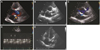
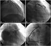
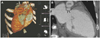
A 42-year-old female was transferred from private clinic with intermittent palpitation and cardiac murmur on July 2012. The patient had no specific medical history of illnesses such as hypertension or diabetes mellitus. There was no family history of aortic, collagen, vascular or congenital heart diseases. Vital signs were blood pressure of 109/64 mm Hg, pulse of 77 beats/min, respiration of 20 breaths/min, and body temperature of 36.5℃. Cardiac auscultation revealed continuous grade 3/6 murmur at the right 2nd and 3rd intercostal spaces. Heart size was normal according to the chest Xray and no apparent ST-segment and T-wave abnormalities were shown on the electrocardiography. The blood chemistries, including N-terminal pro-B natriuretic peptide, coagulation studies with fibrinogen, anti-nuclear antibody and complete blood cell counts, as well as cardiac enzymes and lipid profiles, were within normal limits. TTE and Doppler showed a tunnel-like structure with continuous Doppler signals from the left coronary sinus to the RA (Fig. 1). The pulmonary/systemic blood flow ratio was estimated at 1.3 : 1 measured by echocardiography. The left ventricle showed normal size and systolic function with an ejection fraction of 64%. Cardiac valves were also normal. Exercising treadmill tests showed negative finding. Although coronary angiography revealed the normal left coronary artery (LCA) (Fig. 2A), there was a large tunnel-like structure beginning in the left coronary sinus, and terminating in the body of RA (Fig. 2B and C). The tunnel showed aneurysmal dilatation, and the LCA arose separately from the tunnel (Fig. 2C). Right coronary artery originates from right coronary cusp (Fig. 2D). Cardiac-CT was performed in order to confirm the tunnel, and showed the tunnel taking its origin from the aortic root, passing posterior to the aortic root and entering the RA through a tortuous link (Fig. 3). Although we recommended a surgical treatment, the patient only wanted the surgery next year.
Discussion
Aorta-right atrial tunnel is a rare congenital anomaly characterized by an extracardiac tunnel-like vascular communication arising from an aortic sinus and emptying into the RA. An aneurysmal dilation of the sinus nodal artery has been proposed as the embryologic basis of this unusual lesion. However, the origin of the tunnel from the non-coronary sinus cannot be explained by this theory.1) Other explanations include an abnormal formation of the supravalvular ridge and persistent mesocardial cysts. A more likely cause is a congenital deficiency of the elastic lamina in the aortic media, which gradually enlarges under the influence of high aortic pressure to form an extracardiac tunnel. The preferential anterior or posterior course of the tunnel (from the right or left sinus of Valsalva, respectively) relates to direct anatomic proximity to the low-pressure RA.2) Such aorto-right atrial communication behaves like a left-to-right shunt at the atrial level. Patients with an aorta-right atrial tunnel may be asymptomatic, or they may have exertional breathlessness, palpitations, or recurrent respiratory tract infections.2) It is often detected during the evaluation of a heart murmur in an asymptomatic patient. During periods of increased myocardial oxygen demand, a relatively greater fall in the resistance and subsequent dilation of the coronary arteries prevents coronary steal phenomenon through the tunnel.3) The continued patency of the tunnel poses these risks: calcification of its wall; aortic regurgitation; biventricular volume overload or aneurysmal expansion4); or congestive heart failure, pulmonary vascular disease, infective endocarditis. Two-dimensional echocardiography can differentiate a coronary cameral fistula by visualizing both coronary origins separately away from the tunnel or the tunnel itself.5) Differentiation from a ruptured sinus of Valsalva aneurysm is performed by demonstrating a tunnel with an extracardiac course.5) Coronary angiography is essential to demonstrate the coronary arterial ostia.
Although the need for operative closure in asymptomatic patients remains controversial, surgical closure is recommended after diagnosis because continued patency of the tunnel might predispose patients the risks of volume overload of both ventricles, bacterial endocarditis, aneurysm formation, or spontaneous rupture.6)7) Coil embolization is a useful modality in selective cases. Surgical options include plication of the tunnel or patch closure of aortic origin with direct closure of the atrial opening. If the origin of the coronary artery is deep within the tunnel, it should be re-implanted with a part of the tunnel into the respective sinus of Valsalva.7)
This is the first case report of aorta-right atrial tunnel in Korea detected by TTE, and the previous case was diagnosed by CT.8) The patient had only intermittent palpitation, and she wanted to perform the surgery the following year. So we will follow up on her with TTE after 6 months. In conclusion, the aorta-right atrial tunnel should be included in the clinical differential diagnosis of continuous murmurs which are usually best heard at the right upper sternal border. For the evaluation of chest symptom and murmur, early evaluations with TTE is useful and very important to predict the disease. Then coronary angiography and cardiac CT can be helpful to diagnose the exact disease and they are essential tools for planning the line of treatment.
 XML Download