

 PDF
PDF ePub
ePub Citation
 Print
Print


Introduction
Primary cardiac tumors are rare across all age groups, with a reported prevalence of 0.001% to 0.03% in the autopsy series.1)
Cardiac angiofibroma is a rare cardiac tumor, with only 4 cases having been reported worldwide. Among them, there has been no report that has discussed the tumor's characteristics on the cardiac MRI.
Thus, we report a case regarding an angiofibroma which has primarily originated from the left ventricle of the heart.
Case
A 57 year-old female without any medical history was admitted for a left ventricular (LV) tumor that was discovered incidentally. The tumor was found by echocardiography which was performed for the baseline evaluation of hypertension.
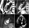
The echocardiography showed a round-shaped, immobile and echogenic mass attached at the LV apex. The valve and LV functions were normal (Fig. 1D).
Cardiac MRI was performed to determine the tumor's tissue type and its relations to other cardiac structures. It showed a slightly high signal intensity on both T1 and T2 weighted images. The gadoliniumenhanced cardiac MRI showed a hypoperfused tumor core and peripheral enhancement (Fig. 1).
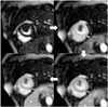
In addition, the lesion showed the centripetal enhancement pattern during the first-pass infusion of the gadolium-containing contrast (Fig. 2). The features of such are associated with the vascular tumors such as liver hemangioma.
The mass showed peripheral enhancement with the central sparing on the delayed enhanced imaging (Fig. 1C). It suggested that the tumor had an abundant fibrous content as well as a vascular content.2)3)
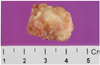
In this patient, the initial preoperative diagnosis of the mass was fibrous tumor of the LV apex. The patient was referred to a cardiac surgeon for the removal of the cardiac mass. Intraoperatively, a whitish, solid mass was detached from the base of the anterolateral papillary muscle (Fig. 3).
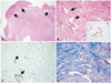
On the histologic evaluation, it showed a somewhat well-demarcated collagenous mass arising from the myocardium (Fig. 4A, arrows). The high-power view showed multiple irregular vascular channels (Fig. 4B) with intervening dense collagen deposition in the mass. The desmin immunohistochemistry and trichrome staining confirmed that the stroma consisted of dense collagenous tissue, not smooth muscle (Fig. 4C and D). From these histologic findings, we diagnosed the mass as angiofibroma. The patient is currently doing well without any evidence of recurrence at the 2-year follow-up.
Discussion
Cardiac angiofibroma is a rare cardiac tumor, with only 4 cases having been reported worldwide. Two of the cases were diagnosed at childhood and related to the systemic congenital defects. In the first case, tuberous sclerosis was associated with the tumor which was found in the child.4) In the second case, Beckwith-Wiedemann syndrome was associated with the tumor.5)
Cao et al.6) reported a case regarding angiofibroma which was located in the right atrium and inferior vena cava. It was unclear whether it was a primary cardiac tumor or vascular tumor extending to the intracardiac cavity. The last case was reported from Russia in 1986.7) None of these cases had the cardiac MRI evaluation; and all 4 cases could not be confirmed as a primary cardiac tumor.
Some tumors, like fibroma, hemangioma and rhabdomyoma, usually arise from the ventricle. Angiofibroma is distinguished from these ventricle-origin tumors by their histologic and image findings such as the cardiac MRI.
In this case, the cardiac angiofibroma showed the centripetal enhancement pattern during the first-pass infusion of the gadolium-containing contrast. The features of such are associated with vascular tumors such as liver hemangioma. It suggested that the tumor had an abundant fibrous content as well as a vascular content.
However, fibromas have higher fibrous content and small vascular content. Therefore, the tumors often demonstrate little or no contrast-material enhancement. Similarly, rhabdomyoma is hypointense to myocardium after the contrast-material administration.
Cardiac hemangioma is a benign cardiac neoplasm which has an abundant vascular component. It shows a more rapid enhancement than angiofibroma during the infusion of the contrast agent.
In immunohistochemistry, CD31 is used primarily to demonstrate the presence of endothelial cells in the histological tissue sections. Desmin is used to demonstrate the smooth muscle cells. In fibroma, the staining for CD 31 and desmin should be negative.
In this case, angiofibroma showed benign vascular proliferations associated with the surrounding collagen deposition. The immunohistochemical staining for CD31 and desmin were positive and negative, respectively.
In summary, we described the MRI and pathologic findings of a cardiac angiofibroma of the LV. These features were considered to be associated with the fibrous and vascular component of the tumor.
 XML Download