

 PDF
PDF ePub
ePub Citation
 Print
Print


Introduction
The presence of anomalous coronary arteries is observed in approximately 1% of patients undergoing coronary angiography (CAG).1) In these patients, the identification of the stenotic ostium and revascularization is difficult, particularly in the emergency setting of primary percutaneous coronary intervention (PCI).
Herein, we report a case of acute myocardial infarction with anomalous separate origin of the left anterior descending artery (LAD) and left circumflex artery (LCX) from the left coronary aortic sinus.
Case
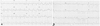
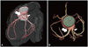
A 59-year-old man with hypertension and type 2 diabetes mellitus visited our emergency room with chest pain that had been started 2 hours prior to his visit. His electrocardiogram showed ST-segment elevation on lead V 1-4 (Fig. 1A). The initial serum Troponin was normal. Total creatine kinase (CK) was elevated to a peak of 4145 Units/L and the CK-MB fraction was 419 ng/mL. An emergency CAG was performed through the right radial artery using a Judkins left catheter. The CAG showed a normal right coronary artery (RCA) and a normal LCX, but no visualization of the LAD. Initially, we considered that the ostium of LAD was totally occluded, but a guide wire could not pass the site through to the LAD ostium (Fig. 2A). On repeated angiography from the coronary sinus, fortunately we found the separate LAD ostium directly from the left coronary aortic sinus beside the LCX ostium without the left main trunk (Fig. 2B and C). There was total occlusion at the proximal LAD. After balloon angioplasty, there was 60% stenosis at the middle LAD (Fig. 2D). We deployed a stent in the proximal LAD. For the demonstration of precise anatomical course and correlation of coronary arteries, coronary computed tomography (CT) angiography was performed with 64-row multidetector CT. It showed an absence of a left main trunk with an anomalous separate origin of LAD and LCX from left coronary aortic sinus (Fig. 3).
Discussion
Coronary artery anomalies are the result of changes that occur during the third week of fetal development and have an overall prevalence from 0.3% to 1.3%. In particular, acute myocardial infarction is a rare clinical presentation in patients with coronary artery anomalies.2)3) Cademartiri et al.4) assessed the prevalence of coronary artery anomalies in patients using coronary CT angiography. In their study, the coronary artery anomaly incidence rates were 86.6% for an RCA anomaly, 9.2% for the left coronary anomaly, and 4.2% for a balanced case. Anomalies of the left coronary are of much lower incidence than those of the right coronary.
In general, the presence of such anomalies does not result in symptoms. Occasionally, more potentially serious anomalies may lead to myocardial ischemia, myocardial infarction, and even sudden cardiac death.5)
Angiographic recognition of unsuspected coronary anomalies is considered important for making an appropriate diagnosis and managing acute myocardial infarction in primary PCI. Repeated failures to identify the anomalous origin of coronary arteries can lead to inadequate diagnosis and prolonged procedures, which can result in serious complications.6) Although their incidence is low, clinicians should always take into consideration coronary artery anomalies.
Coronary CT angiography is particularly useful to clarify the relationship between coronary arteries and great vessels and the correct position of coronary arteries ostia. Currently, the ideal imaging tool for the diagnosis and delineation of coronary artery anomalies is coronary CT angiography.7) In this case, it was very difficult to find the ostium of the obstructed anomalous LAD during primary PCI. Coronary CT angiography precisely demonstrated a separate origin of LAD and LCX from the left coronary aortic sinus after PCI.

 XML Download