

 PDF
PDF ePub
ePub Citation
 Print
Print


A 58-year-old man with paroxysmal supraventricular tachycardia was admitted to our hospital for catheter ablation.
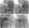
Cannulation of the patient's coronary sinus using a long sheath multipolar catheter was difficult and the catheter was positioned in the interatrial septum. The tip of the catheter was directed to the left atrium (Fig. 1A). Angiography using the long sheath catheter showed a vein (Fig. 1B, arrows) draining to the left atrial appendage (Fig. 1B, arrowheads). The septal vein of the left atrium could be visualized in the right anterior oblique (Fig. 1C) and left anterior oblique views (Fig. 1D) after placement of a multipolar catheter in the coronary sinus. Atrioventricular nodal reentrant tachycardia was induced by programmed electrical stimulation and slow pathway modification was performed successfully.
Difficult cannulation of the coronary sinus may be due to Thebesian valve, valve of Vieussens or congenital anomaly such as atresia of the coronary sinus ostium.1)
Little attention has been paid to the atrial veins, except vein of Marshall as an arrhythmogenic focus.2)
There are 3 groups of veins draining the left atrium: septal veins, lateral veins and proper veins of the left atrium.3) The veins from the left atrial appendage are drained usually via the lateral atrial vein into the coronary sinus. In this case, the vein from the left atrial appendage was connected to the septal vein.
In conclusion, we report incidental angiographic visualization of an interatrial septal vein during a cardiac electrophysiology study.
 XML Download