

 PDF
PDF ePub
ePub Citation
 Print
Print


Introduction
Atherosclerosis is the leading cause of peripheral artery disease (PAD) and coronary artery disease (CAD). Some previous studies have reported that PAD is a coronary heart disease risk equivalent.1)2) Another study, however, reported that risk factors for PAD are different to those for CAD.3) Risk factors for atherosclerosis are hypertension (HT), diabetes mellitus (DM), hypercholesterolemia {high total cholesterol (TC) levels, high low density lipoprotein-cholesterol (LDL-C) levels, and/or low high density lipoprotein-cholesterol (HDL-C) levels}, smoking, older age, obesity, metabolic syndrome (MetS)4) and chronic kidney disease (CKD).5) Risk factors for PAD may vary depending on the affected arteries.6)
Although the prevalence of PAD and CAD is known to increase with the ageing population,7) few studies have compared risk factors for PAD to CAD solely in the Korean population. Therefore, our objective in the present study was to analyze and compare risk factors for PAD, CAD, and for normal controls under the hypothesis that risk factors for PAD and CAD are different from those for normal controls.
Subjects and Methods
Study population and design
We reviewed the records of patients diagnosed with PAD and CAD at the Cardiac and Vascular Center from November 1994 to November 2004 as well as those of healthy subjects (normal control group; Control) who underwent health examinations of digestive organs during the same period at the Health Promotion Center of Samsung Medical Center. We excluded patients with cardiovascular disease (CVD), cerebrovascular accident, or lung cancer from the normal control group. The enrolled subjects consisted of 1) patients with PAD (n=415) who had over 50% peripheral artery occlusion confirmed by lower extremity computed tomography angiography, 2) patients with CAD (n=3686) including those with stable angina, unstable angina, and acute myocardial infarction confirmed by cardiac catheterization, and 3) Control (n=3835). In addition, self reported information on the absence of CAD and PAD was used in control. Information was obtained by reviewing electronic medical charts. This study was approved by the Samsung Medical Center institutional review board; informed consent was waived for this retrospective study.
Diagnostic criteria
Cardiovascular risk factors
Subjects were defined as having HT if they were taking an anti-HT drug, had been clinically diagnosed with HT, or had either a systolic blood pressure (SBP) ≥40 mm Hg or a diastolic blood pressure (DBP) ≥90 mm Hg. Subjects who met one of the following requirements were defined as having DM: on an oral hyperglycemic agent, using insulin, clinical diagnosis of diabetes, or a fasting glucose level >126 mg/dL. Subjects were defined to have hypercholesterolemia if they met one of the following requirements: diagnosis of hypercholesterolemia or a medication history of hypercholesterolemia or TC >200 mg/dL or LDL-C >130 mg/dL. The following body mass index (BMI) categories were recognized: normal (18.5≤BMI<23), overweight (23≤BMI<25) and obese (BMI≥25). There was no statistical meaning of adding an underweight category because the number of underweight patients was two in the PAD group, thus we included them into the BMI normal group in PAD. A patient who had smoked within a year prior to the study was defined as a smoker. The estimated Glomerular Filtration Rate (eGFR), which was used as an indicator of kidney function, was calculated using the Modification of Diet Renal Disease Study formula: eGFR (mL/min/1.73 m2)=186.3×{serum creatinine (Cr)}-1.154×(age)-0.203×(0.742 if women)×(1.21 if African-Americans).
The National Kidney Foundation Kidney Disease Outcome Quality Initiative defined CKD as an eGFR <60 mL/min/1.73 m2. Patients with MetS were classified into two groups based on the modifications suggested by the National Cholesterol Education Program Adult Treatment Panel III.1) Diagnosis of MetS in this study was based on the presence of three or more of the following symptoms: 1) BMI ≥25 (BMI categories for Asia of International Obesity Taskforce), 2) triglyceride (TG) levels ≥150 mg/dL, 3) HDL-C levels <40 mg/dL for men and <50 mg/dL for women, 4) HT with SBP ≥130 mm Hg, DBP ≥85 mm Hg, or undergoing active antihypertensive drug therapy, and 5) fasting blood sugar (FBS) ≥100 mg/dL or active use of oral hypoglycemic agents or insulin.
A number of PAD patients had had CAD, however, CAD patients were not diagnosed with PAD in our data. Patients with PAD were divided into two groups based on the absence or presence of coexisting CAD. PAD subjects were also classified into two groups based on the modified recommendations of Haltmayer et al.8) according to the affected arteries of the lower limb: 1) aortoiliac (AI) disease including occlusion or >50% stenosis in the abdominal aorta and common and external iliac arteries, 2) femoropopliteal (FP) disease including occlusion or >50% stenosis in the common, superficial and deep femoral, popliteal, and infrapopliteal arteries. The FP and AI groups were compared with the CAD group, because the coronary arteries are similar to the FP arteries in size.
Statistical analysis
General characteristics of PAD, CAD, and Control subjects were analyzed by one-way analysis of variance with the Bonferroni method in multiple comparisons testing for continuous variables. The χ2-test was used to compare categorical variables. To analyze and compare risk factors between PAD subjects with coexisting CAD and those with no coexisting CAD and between PAD subjects with AI and FP, we employed Student's t-test for continuous variables and the χ2-test for categorical variables. Simple logistic regression analysis and multinomial logistic regression analysis were carried out to determine the association among cardiovascular risk factors in PAD, CAD, and control subjects. Two models were used to adjust variables. In Model I, age, gender, HT, DM, hypercholesterolemia, obesity grade (obese), smoking status and CKD were adjusted. Model II considered age, gender, smoking status, CKD, and MetS.
Results
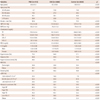
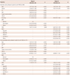
The mean age of PAD subjects was 64.4 (±9.3) years, while the mean age of CAD subjects was 61.2 (±9.9) years, and that of Control subjects was 59.9 (±9.1) years (p<0.001). The proportion of males in the subject groups was as follows: 90.6% for PAD, 71.4% for CAD, and 75.5% for Control (p<0.001). More PAD subjects than CAD and Control subjects had HT, DM, and CKD (p<0.001), while more CAD subjects were smokers, had hypercholesterolemia, and were obese than PAD and Control subjects (p<0.001). Among the components of MetS, more PAD subjects had high blood pressure and high FBS (p<0.01) than patients in the other two groups, while more CAD subjects had low HDL-C levels and were obese than PAD and Control subjects (p<0.01). TC, TG, LDL-C, HDL-C, FBS, and Cr were significantly different among the three groups (p<0.001). The results after Bonferroni correction for multiple comparisons are shown in Table 1.
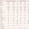
Among PAD subjects, the proportion of AI subjects with coexisting CAD was 58.4%, while the proportion of PAD patients with no coexisting CAD was 56.8% {p=nonsignificant (NS)}. The proportion of coexisting CAD was 33.6% in PAD patients with AI and 32.2% in PAD patients with FP (p=NS) (Table 2).
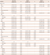
In Model I, the adjusted odds ratios (ORs) for HT {OR 6.43, 95% confidence interval (CI) 4.92-8.39}, DM (OR 7.71, 95% CI 6.05-9.84), hypercholesterolemia (OR 1.50, 95% CI 1.18-1.90), smoking (OR 10.3, 95% CI 7.89-13.4), and CKD (OR 1.54, 95% CI 1.14-2.04) were significantly higher in subjects with PAD compared to those in normal controls. However, the ORs for HDL-C (OR 0.92, 95% CI 0.91-0.93), being overweight (OR 0.51, 95% CI 0.38-0.67), and being obese (OR 0.32, 95% CI 0.24-0.43) were significantly lower in PAD subjects compared to those in normal controls. The ORs for HT (OR 2.89, 95% CI 2.57-3.25), DM (OR 4.90, 95% CI 4.28-5.61), smoking (OR 3.76, 95% CI 3.28-4.30), being overweight (OR 1.21, 95% CI 1.04-1.41), being obese (OR 1.74, 95% CI 1.51-2.01) and CKD (OR 1.46, 95% CI 1.24-1.72) were significantly higher in CAD subjects than those in normal controls. However, the ORs for hypercholesterolemia (OR 0.67, 95% CI 0.60-0.76) and HDL-C (OR 0.93, 95% CI 0.92-0.94) were significantly lower in the CAD group than those in the control group.
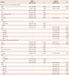
In Model II, the ORs for smoking (OR 9.38, 95% CI 7.35-11.9), CKD (OR 2.14, 95% CI 1.63-2.82), and MetS (OR 3.61, 95% CI 2.89-4.51) were higher in PAD subjects than those in normal controls. Comparing CAD subjects with normal control group subjects, the ORs for smoking (OR 3.36, 95% CI 2.99-3.79), CKD (OR 1.80, 95% CI 1.56-2.09), and MetS (OR 3.66, 95% CI 3.31-4.05) were significantly higher in CAD subjects (Table 3).
We analyzed the association between cardiovascular risk factors in 1) PAD patients with or without coexisting CAD, and in the normal control group and 2) the PAD affected site; AI or FP, and normal control group. The ORs in PAD patients with or without coexisting CAD group and AI or FP group for HT, DM, smoking, HDL-C, and obese grade (Model I) and smoking, MetS, and CKD (Model II) were similar to PAD subjects (Table 4 and 5).
Discussion
The overall findings of this study revealed that HT, DM, hypercholesterolemia, obesity, smoking, CKD and MetS are risk factors for PAD and CAD. However, obesity as a risk factor showed inconsistent results between PAD and CAD.
These findings are consistent with those of previous studies that reported that risk factors for PAD are similar to those for CAD. For instance, HT,9) DM,10) hypercholesterolemia,11) smoking,12) CKD,13) and MetS4) are known risk factors for CAD. Furthermore, HT,14) DM,15) hypercholesterolemia,16) smoking,17) CKD,5) and MetS4) are risk factors for PAD. In our study, the mean TC, LDL-C, HDL-C values were higher in the normal control group than those in the PAD or CAD group. The results from a Japanese male worker study,18) a Korean study,19) and a U.S. adult study based on the National Health and Nutrition Examination Surveys20) are consistent with the high TC, LDL-C, HDL-C values that we found in our normal control group.
Obesity is one of the major risk factor for CVD, including PAD.21) Obesity is also associated with high mortality related to chronic disease.22) However, patients with CAD or PAD have an inverse correlation between BMI and cardiovascular mortality after adjustment for confounding variables in the Factores de Riesgo y ENfermedad Arterial registry.23) The obesity paradox24) is that obese patients receive better treatment and care, because they are perceived to be at high risk. However, our PAD obesity results cannot be explained by the obesity paradox, because PAD subjects had occlusion or >50% stenosis in the peripheral artery confirmed by lower extremity computed tomography angiography. This corresponds to an ankle-brachial index <0.9 and the consequent development of intermittent claudication, gangrene, and pain. This leads to physically and mentally instable condition, leading to weight loss. Our PAD subjects who visited tertiary medical services already had progressed PAD with a limited radius of action, immobilization, muscle atrophy, and depression requiring surgery or intervention. The factors outlined above may explain why the obesity results were inconsistent between PAD and CAD subjects in our analysis.
The ORs for the risk factors for the absence or presence of coexisting CAD in PAD and CAD, with the exception of obesity, were similar among the groups. A study on the association of cardiovascular risk factors with patterns of lower limb atherosclerosis in 2659 patients who underwent angioplasty revealed that DM predicted PAD compared to no DM or current smoking status.25) An Italian study also showed that preexisting CAD with PAD was associated with risk factors for PAD.26) In our study, 33% of subjects with PAD had coexisting CAD. This result is consistent with previous studies that reported 21% of PAD subjects showed myocardial infarction and 26% of PAD subjects had angina.27) A comparison of the ORs for AI and FP according to the affected site in PAD and CAD revealed similar risks as those reported in a Turkish study (Ankara),28) and two U.S.A-based studies (San Diego29) and southern California30)).
This study had several limitations. First, the study was conducted retrospectively at a single center, which may have caused selection bias. We were also not able to eliminate the possibility of information bias when collecting medical records from the medical charts of the subjects and laboratory results. Second, the mean age of the subjects in the normal control group was younger than that of the subjects in the PAD and CAD groups. To minimize the effect of age, we conducted age-adjusted analysis. A further limitation of our study is that we could not consider symptoms of patients with PAD, physical activity, nutrition, socioeconomic position, waist circumference, or health behavior variables due to limited data. Further cardiovascular cohort studies considering these variables are therefore required to verify the risk factors for atherosclerosis.
Conclusions
We found significantly different ORs for risk factors, namely age, gender, HT, DM, hypercholesterolemia, HDL-C, obesity, smoking, CKD, and MetS, in the PAD and CAD groups compared to those in the Control group. Interestingly, the ORs for obesity were inconsistent between PAD and CAD subjects. In other words, obesity grade was showed opposite trends. However, in both diseases, cardiovascular risk factors were found to be risk factors. In conclusion, there appears to be no differences in risk factors for PAD and CAD in the Korean population.
 XML Download