Abstract
Percutaneous recanalization of chronic total occlusions (CTOs) in peripheral arteries, especially TASC D classification including the distal aorta and both iliac arteries is still technically challenging. The conventional technique using standard guidewires and catheters guided by computed tomography and angiography can achieve a limited initial success, depending on lesion characteristics and operator's experience. A special imaging technique using 3-dimensional rotational angiography and spatio-temporal reconstruction with endoview for a better examination of the proximal stump, exact obstruction location, and distal stump direction in a stumpless lesion can be indispensable for successful intervention. We report a successful revascularization case of stumpless distal aorta and bi-iliac CTO guided by this specialized imaging technique.
Most patients with peripheral artery chronic total occlusions (CTOs) suffer from critical limb ischemia or disabling claudication.1) Recanalization of CTOs in peripheral arteries is still technically and clinically challenging due to a relatively limited procedural success rate and higher complication rate. The conventional technique using standard guide wires and catheters can achieve an initial success rate in about 40-60% of cases,2) depending on lesion characteristics (length, location, calcification and runoff vessel status) and operator's experience. A significant proportion of procedural failure has resulted from limited anatomical information with conventional imaging tools.3) Thus, additional detailed anatomical evaluation by newer imaging techniques may play an important role to obtain further information regarding exact obstruction location, severity of stenosis and accurate indications such as microchannels in the case of a stumpless lesion. Although conventional CT angiography and invasive angiography are useful in usual patients with CTOs, in particular cases involving the stumpless ostial lesion, CT and invasive angiography are not enough for detecting accurately the stump of an occlusive lesion and associations to the surrounding vessel architecture. Three-dimensional (3D) rotational angiography is an image acquisition technique that displays vascular structures in a 3D like format. This technique provides significantly more visual information regarding stump location, the real vessel length and lumen diameter of measurements than conventional angiography.4)
In this case, we successfully treated a patient with a stumpless total occlusion at the distal aorta and bi-iliac occlusion guided by 3D rotational angiography and spatio-temporal reconstruction.
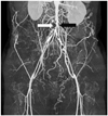
A 54-year-old man presented with severe claudication and coldness in both lower extremities several years ago. The ankle-brachial index of both lower extremities was not measurable. The lower extremity CT angiogram showed total occlusion from the infra-renal distal abdominal aorta to both external iliac arteries. There was a2013-05-06 well-developed inferior mesenteric artery (IMA) forming multiple collaterals at the distal abdominal aorta, but a definite angiographic stump of the distal aorta to the iliac arteries was not visible (Fig. 1). A CT angiogram was not enough to detect the stump of an occlusive lesion in the distal aorta and relationships to surrounding vessels. So we considered 3D full rotational angiography and spatio-temporal reconstruction for detecting the stump at the distal aorta and additional vascular structures.
First, an aortography was performed via radial approach with a 5 Fr pigtail catheter. The aortography showed that there was total occlusion at the distal abdominal aorta below both renal arteries and total occlusion at the proximal bi-iliac arteries. Although there was a severe discrete concentric stenosis in the ostium (OS) of IMA, the entire vessel was well-developed which gave rise to profuse collaterals to the bi-iliac arteries from the distal to the occluded segment.
Vascular access was achieved via both femoral arteries and right brachial artery. 8 Fr short sheaths were inserted to both femoral arteries for a retrograde approach and an 8 Fr Vistabrite IG guiding sheath (Johnson & Johnson, Miami, FL, USA) was inserted to the right brachial artery for anterograde approach.
A full rotational 3D angiography was performed and spatiotemporal reconstruction was carried out by Philips cine angiography FD 20 software (Philips, Amsterdam, the Netherlands). Mapping with 3D reconstructed images, an endoview showed a blunt stump in the distal aorta just distal to the IMA OS which was not shown on the CT or invasive diagnostic aortography (Fig. 2).
There was a suspicious micro-channel from the distal abdominal aorta to the right iliac artery from 3D rotational angiography (Fig. 2, transparent arrow). This micro-channel was not observed by previous CT angiography. A virtual line could be drawn from the suspicious stump in the distal abdominal aorta to the right iliac artery (Fig. 2, dot line). A successful guidewire passage was achieved through this micro-channel.
Under the 3D image guide, aggressive bilateral kissing wiring was performed using 035 soft long Terumo wire (0.035 Radiofocus® Guidewire M, Terumo Corp., Somerset, NJ, USA) under the 5 Fr multipurpose-1 catheter support from the distal aorta to the right iliac artery and from the right femoral artery to the right iliac artery by subintimal approach (Fig. 3A). After successful 035 guidewire passage from the aorta to both iliac arteries, sequential predilation was done using Powerflex 6.0×80 mm (Johnson & Johnson, Miami, FL, USA) from the distal aorta to both external iliac arteries (Fig. 3B). After the predilation of both entire iliac arteries to the distal aorta, simultaneous kissing stenting was performed using two Smart control stents {7.0×100 mm (aorta to right iliac artery)/6.0×100 mm (aorta to left iliac artery) (Fig. 3C)}. Residual stenosis was approximately 40% and good distal flow was observed. To get optimal angiographic results, final kissing ballooning was carried out using two Powerflex 6.0×80 mm balloons. A final angiogram showed excellent angiographic outcomes (good distal run-off without flow limitation) (Fig. 3D). The patient was stabilized and safely discharged for regular clinical follow up.
Recanalization of CTOs is posing a big challenge in achieving procedural success by its complex nature. Peripheral interventionists now have several technologies available to address the challenges of crossing CTO lesions and re-entering the distal true lumen.
Kawasaki et al.5) described a new technique for the treatment of iliac and femoropopliteal occlusions, in which intravascular ultrasound is used to guide a stiff guidewire to cross the true lumen of CTO lesions. Kawarada et al.6) described the practical use of duplex echo-guided recanalization of CTO in the iliac artery. Dvir et al.7) described that 3D reconstruction of CTOs can clearly image the stump area, delineate the lesion path, and provide enough information for the clinician to precisely calculate the severity of stenosis and lesion length.
To our knowledge, there is no 3D angiography and reconstructed image guided recanalization of CTO in the distal aorta and both iliac arteries. Clearly, there is a need to determine the best approach by an optimal image guide to improve technical success for lower extremity CTO recanalization.
The present case report is a typical case in which demonstrated successful revascularization by 3D rotational angiography and 3D reconstruction for the stumpless long CTO lesions from the distal aorta to the bi-iliac arteries. This particular 3D reconstruction and subsequent spatiotemporal understanding from the exoview (the usual luminogram showing in the conventional CT angiography or invasive angiography) and endoview enabled us to detect clues for the stumpless CTO lesion. Moreover, we could obtain more detailed information regarding the location, direction, size and relationship with adjacent side branch (IMA) of the blunt stump through endoview. Further, we could detect a suspicious micro-channel from the distal abdominal aorta to the iliac artery by endoview following 3D reconstruction, which was not seen by previous CT angiography. Finally, successful wiring through this micro-channel was done and procedural success was achieved. The present case demonstrates that we need more additional structural and anatomical information through more specific images provided by 3D rotational angiography to find out definite clues for the stumpless CTO lesions.
With 3-dimensional rotational angiography, we might have used more contrast and been exposed to more radiation due to prolonged contrast injection and prolonged cine angiography to get more clean images. Thus, we should consider the baseline renal function of a patient and should confront prolonged procedure time with more radiation exposure.8) Consequently we should choose patients selectively for 3D rotational angiography guided revascularization. We would like to suggest that the usage of 3D rotational angiography and spatiotemporal reconstruction may serve as a useful tool for planning interventional procedures for stumpless peripheral CTO lesions and improving their success rate.
Figures and Tables
Fig. 1
Preprocedural CT angiographic findings. Preprocedural CT angiography showed that there was total occlusion from the distal aorta to both iliac arteries. Inferior mesenteric artery from the distal abdominal aorta (black arrow) was hypertrophied and prominent. But there was no angiographically visible stump in the distal aorta at the inferior mesenteric artery ostium level (white arrow).

Fig. 2
Invasive 3D angiographic image of pre-stenting. A: 3D angiographic image. B: endoview at the distal aorta total occlusion level from 3D reconstruction image. A virtual line was drawn from the distal abdominal aorta to right iliac artery (dot line). There was a suspicious micro-channel from the distal abdominal aorta to right iliac artery (transparent arrow). This micro-channel was not seen by previous CT angiography. There was a visible inferior mesenteric artery from distal aorta (black arrow). 3D: three-dimensional.

Fig. 3
Successful complete revascularization and the final results. A: bilateral kissing wiring using 035 soft Terumo wires under the 5 Fr multipurpose catheter support by subintimal tracking from distal aorta to external iliac artery. B: predilation in right iliac artery after successful kissing wiring. C: bilateral kissing stenting using two self-expanding nitinol stents from distal aorta to both iliac arteries. D: postprocedural angiography.

References
1. Gallagher KA, Meltzer AJ, Ravin RA, et al. Endovascular management as first therapy for chronic total occlusion of the lower extremity arteries: comparison of balloon angioplasty, stenting, and directional atherectomy. J Endovasc Ther. 2011. 18:624–637.
2. van der Heijden FH, Eikelboom BC, Banga JD, Mali WP. Management of superficial femoral artery occlusive disease. Br J Surg. 1993. 80:959–963.
3. Al-Ameri H, Shin V, Mayeda GS, et al. Peripheral chronic total occlusions treated with subintimal angioplasty and a true lumen re-entry device. J Invasive Cardiol. 2009. 21:468–472.
4. Lee JB, Chang SG, Kim SY, et al. Assessment of three dimensional quantitative coronary analysis by using rotational angiography for measurement of vessel length and diameter. Int J Cardiovasc Imaging. 2012. 28:1627–1634.
5. Kawasaki D, Tsujino T, Fujii K, Masutani M, Ohyanagi M, Masuyama T. Novel use of ultrasound guidance for recanalization of iliac, femoral, and popliteal arteries. Catheter Cardiovasc Interv. 2008. 71:727–733.
6. Kawarada O, Yokoi Y, Takemoto K. Practical use of duplex echo-guided recanalization of chronic total occlusion in the iliac artery. J Vasc Surg. 2010. 52:475–478.
7. Dvir D, Assali A, Kornowski R. Percutaneous coronary intervention for chronic total occlusion: novel 3-dimensional imaging and quantitative analysis. Catheter Cardiovasc Interv. 2008. 71:784–789.
8. Pannu N, Wiebe N, Tonelli M. Alberta Kidney Disease Network. Prophylaxis strategies for contrast-induced nephropathy. JAMA. 2006. 295:2765–2779.


 PDF
PDF ePub
ePub Citation
 Print
Print


 XML Download