

 PDF
PDF ePub
ePub Citation
 Print
Print


Abstract
Background and Objectives
Hypercholesterolemia is a key factor in the development of atherosclerosis. We sought to evaluate the relation between hypercholesterolemia and plaque composition in patients with coronary artery disease.
Subjects and Methods
Study subjects consisted of 323 patients (mean 61.5 years, 226 males) who underwent coronary angiography and virtual histology-intravascular ultrasound examination. Patients were divided into two groups according to total cholesterol level: hypercholesterolemic group (≥200 mg/dL, n=114) and normocholesterolemic group (<200 mg/dL, n=209).
Results
Hypercholesterolemic patients were younger (59.7±13.3 years vs. 62.6±11.5 years, p=0.036), than normocholesterolemic patients, whereas there were no significant differences in other demographics. Hypercholesterolemic patients had higher corrected necrotic core volume (1.23±0.85 mm3/mm vs. 1.02±0.80 mm3/mm, p=0.029) as well as percent necrotic core volume (20.5±8.5% vs. 18.0±9.2%, p=0.016) than normocholesterolemic patients. At the minimal lumen area site, percent necrotic core area (21.4±10.5% vs. 18.4±11.3%, p=0.019) and necrotic core area (1.63±1.09 mm2 vs. 1.40±1.20 mm2, p=0.088) were also higher than normocholesterolemic patients. Multivariate linear regression analysis showed that total cholesterol level was an independent factor of percent necrotic core volume in the culprit lesion after being adjusted with age, high density lipoprotein-cholesterol , hypertension, diabetes mellitus, smoking and acute coronary syndrome (beta 0.027, 95% confidence interval 0.02-0.053, p=0.037).
Figures and Tables
Fig. 1
Representative VH-IVUS findings in both groups. A: normocholesterolemic patient (42 years old man with stable angina pectoris) showed small amounts of necrotic core volume. B: hypercholesterolemic patient (55 years old man with ST-segment elevation myocardial infarction) showed large amounts of necrotic core volume. VH-IVUS: virtual histology-intravascular ultrasound, EEM: external elastic membrane, CSA: cross sectional area.
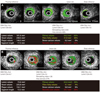
Table 1
Clinical characteristics of the study subjects
MI: myocardial infarction, STEMI: ST-segment elevation myocardial infarction, LAD: left anterior descending artery, LCX: left circumflex artery, RCA: right coronary artery, LM: left main artery, HDL-C: high density lipoprotein-cholesterol, LDL-C: low density lipoprotein-cholesterol, FBS: fasting blood sugar, BUN: blood urea nitrogen, hs-CRP: high sensitivity C-reactive protein
Table 2
Virtual-histology intravascular ultrasound finding
 XML Download