Journal List > Korean Circ J > v.43(3) > 1016999

Jo, Kim, Kim, Kim, Han, Park, and Choi: Evaluation of Short-Term Use of N-Acetylcysteine as a Strategy for Prevention of Anthracycline-Induced Cardiomyopathy: EPOCH Trial - A Prospective Randomized Study

Abstract

Background and Objectives

We investigate to determine whether N-acetylcysteine (NAC) can prevent anthracycline-induced cardiotoxicity.

Subjects and Methods

A total of 103 patients were enrolled in this prospective randomized open label controlled trial. They are patients first diagnosed with breast cancer or lymphoma, who require chemotherapy, including anthracycline like adriamycine or epirubicine. Patients were randomized to the NAC group {n=50; 1200 mg orally every 8 hours starting before and ending after the intravenous infusion of anthracycline in all chemotherapy cycles (3-6)} or the control group (n=53). Primary outcome was the decrease in left ventricular ejection fraction (LVEF) absolutely ≥10% from the baseline and concomitantly <50% at 6-month. Composite of all-cause death, heart failure and readmission were compared.

Results

The primary outcome was not significantly different in the NAC and control groups {3/47 (6.4%) vs. 1/52 (1.9%), p=0.343}. The mean LVEF significantly decreased in both the NAC (from 64.5 to 60.8%, p=0.001) and control groups (from 64.1 to 61.3%, p<0.001) after the completion of whole chemotherapy. The mean LVEF change did not differ between the two groups (-3.64% in NAC vs. -2.78% in control group, p=0.502). Left ventricular (LV) end systolic dimension increased with higher trend in NAC by 3.08±4.56 mm as compared with 1.47±1.83 mm in the control group (p=0.064). LV end diastolic dimension did not change in each group and change does not differ in both. Peak E, A and E/A ratio change and cardiac enzymes were comparable in two groups. Cumulative 12-month event rate was 6% and 3.8% in the NAC group and the control group, respectively, with no difference (p=0.672).

Conclusion

We cannot prove that NAC prevents anthracycline-induced cardiomyopathy.

Introduction

A growing population are diagnosed with cancers and treated with chemotherapeutic agents. Among them, anthracyclines are highly effective at treating certain cancers. However, their use is limited by the potential for cardiotoxicity. Several studies address a wide range of the incidence of cardiotoxicity, which is related to differences in the definitions, chemotherapy regimens, and patient populations. The occurrence of clinical heart failure (HF) seems to be between 1% and 5%, and asymptomatic decrease in the left ventricular function is in the range of 5% to 20%.1)2)
Recent development in the field of oncology, including early diagnosis and new anti-cancer-drugs, have increased the survival of cancer patients and accordingly, be more likely to have cardiac damage, as like HF by cardiotoxic chemotherapy. Moreover, patients with cancers are mostly older, and likely to have concomitant ischemic heart disease. This confers them more vulnerable to cardiac insult from cardiotoxic chemotherapeutic agents. HF induced by chemotherapeutic agents may compromise the clinical effectiveness of chemotherapy, affecting the patient's survival and quality of life, independently of the prognosis related to the cancer per se.
The representative cardiac toxic chemotherapeutic agents are anthracycline (ANT), including adriamycine, epirubicine and daunorubicine, which are mainly used in patients with breast cancer, lymphoma and sarcoma. In view of the vastly growing number of patients with breast cancer in Korea, who will most likely receive chemotherapy, including ANT, it is necessary to look for strategy of managing or preventing chemotherapy induced HF. When we consider that ANT-induced cardiomyopathy is irreversible and seems to have a similar impact on survival as the other forms of HF, the prevention is mandatory.3)
Although the mechanism of ANT-induced cardiomyopathy is not fully elucidated, the oxidative stress is believed to be most plausible4-8) among many suggested ones, including apoptosis, elevated calcium accumulation in mitochondria, and modulation of cardiac gene expression.4-6)
There have been reports on the potential benefit of N-acetylcysteine (NAC), a representative antioxidant, in preventing doxorubicin cardiac toxicity in mouse models.9) There has been, however, no clinical trial to date dealing with humans.
We investigate a short-term high dose NAC as a potential drug for the prevention of ANT induced cardiomyopathy. This is the first clinical trial to reappraise the potential of NAC as preventive strategy for preventing chemotherapy induced cardiac toxicity.

Subjects and Methods

The "Evaluation of Short-Term Use of N-acetylcysteine as a Strategy for Prevention of Anthracycline-Induced Cardiomyopathy (EPOCH)" trial was conducted in Hallym University Sacred Heart Hospital, in Korea from June 2008 to December 2010. A total of 103 patients, who meet the inclusion criteria, were allocated to each study arm. All the patients have breast cancer or lymphoma and receive ANT based chemotherapy as doxorubicin or epirubicine.
The inclusion criteria were as followings: patients with only right breast cancer who can protect the heart from radiation therapy on the breast. Patients were first diagnosed with cancer at this time and received chemotherapy, 19 years old or more, the baseline left ventricular ejection fraction (LVEF) ≥50% and patients who agreed to an informed consent.
Exclusion criteria are as follows: the baseline LVEF <50%, patients with HF, patients with dilated cardiomyopathy or restrictive cardiomyopathy, moderate or severe aortic or mitral valve insufficiency, patients who were previously diagnosed with cancer, patients who received previous chemotherapy or radiation therapy at any organ at any time, patients with left-sided breast cancer, aspartate aminotransferase (AST)/alanine aminotransferase (ALT) 3-times or more of upper limits, creatinine kinase (CK) over upper normal limits, patients who received other antioxidant as vitamin C, E or probucol within 3-month before chemotherapy and life expectancy of less than a year.

Primary endpoint

The primary endpoints of EPOCH trial is the decrease of LVEF absolutely 10% or more and concomitant reduction below 50% at 6-month follow-up echocardiography, after the completion of whole cycles of chemotherapy.

Secondary endpoints

The secondary endpoints are LVEF decrease of absolute value of 10% or more, peak CK, CK-MB and cTnI at the end of each cycle of chemotherapy, and LVEF and Doppler parameters as peak E, A, E/A at 6-month after the completion of chemotherapy measured with echocardiography. Clinical outcomes, including all cause mortality, HF, and re-hospitalization, were also compared at 1-, 3-, 6- and 12-months after the whole cycles of chemotherapy.

Randomization

Eligible patients were randomly assigned to receive NAC or placebo. Patients were randomized 1 : 1, by a computer-generated permuted block of 6 patients to ensure the balanced assignment of patients to each group. Randomization codes were provided by the research nurse at Hallym University Sacred Heart Hospital Cardiovascular Center.

Study protocol

After identification of the study candidates, informed consent was obtained and eligible study patients were randomized. Patients randomized to the NAC group received 1200 mg every 8 hours, for 2 days: thrice before ANT administration, with the last dosing just 1 hour before ANT administration, because the best results were attained from the previous animal study with that schedule;9) and thrice after ANT administration, beginning 8-hours after the chemotherapy in every cycle. A total of 7200 mg of NAC was administered with six 1200 mg doses per cycle.
Adriamycine was injected with one bolus through the peripheral venous route, and epirubicine was also administered intravenously over 3-5 minutes. Other combined chemotherapeutic agents are administered by routine protocols of chemotherapy. This protocol was repeated in every 4-6 cycles. Therefore, the total dose of NAC ranges from 28800 to 43200 mg through the whole chemotherapy.
All study patients underwent a baseline laboratory test, including cardiac markers and echocardiography. LV systolic function, LV chamber size and mitral inflow pattern are measured within a week before the 1st cycle of chemotherapy. The cardiac markers as CK, CK-MB, and cTnI were measured per 24 hours in each chemotherapy cycle, for 2 days after each ANT administered, and echocardiographic follow up at 6-month after last chemotherapy was also performed.
The clinical outcomes, including all cause death, HF, and unscheduled re-hospitalization, were also compared at 1-, 3-, 6- and 12-month after the last chemotherapy.

Sample size calculation and statistics

The primary endpoint is the decrease of LVEF ≥absolute value of 10% from the baseline and concomitantly decline below 50% reflecting the definition of chemotherapy induced cardiotoxicity from international oncological guidelines.10)11)
We assumed patients who received NAC will attain significantly lower primary outcome, as compared to the control, 10% in the NAC treatment group and 35% in the control group, referring to the similar clinical trial using enalapril, as a study drug.12) Therefore, the difference is 25% between the 2 groups. Using this assumption, the study population of 43 patients per group would permit a 2-sided significance level of 5% and 80% power. To allow for the possibility of patients lost during follow-up, incomplete data collection, and protocol violations (20%), the planned sample size was 54 patients in each group.
The baseline demographic and procedure-related characteristics were analyzed from the intention-to-treat population. Laboratory data, including follow-up echocardiography parameter measurements, were analyzed from the per-protocol population. Continuous variables were expressed as the means and standard deviations, while categorical variables were expressed as absolute numbers and proportions of patients in a given category. Data were compared using the chi-square test or Fisher's exact test (categorical variables) and the Student t-test (continuous variables).

Results

Patients' demographics and disposition

Between June 2008 and December 2010, 103 were randomized to receive either NAC (n=50) or control (n=53). After exclusions, due to protocol violations, incomplete laboratory test results, and insufficient follow-up, 47 patients receiving NAC and 52 patients in control were included in the per-protocol analysis. Cancer type, chemotherapeutic regimen, total dose, and number of cycles were comparable. The demographic and baseline data are provided in Table 1. The patient groups did not differ significantly.

Primary endpoint

Three patients in the NAC group (6.4%) and 1 patient in the control group (1.9%) had a decrease of LVEF by absolute value of ≥10% and below 50% at the 6-month follow-up echocardiography, after the completion of whole cycles of chemotherapy, including ANT (p=0.343) (Fig. 1).
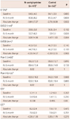
The mean LVEF significantly decreased in both groups; from 64.5 to 60.8% (p=0.001) in the NAC group and from 64.1 to 61.3% (p<0.001) in the control group after chemotherapy. The mean absolute LVEF change was not different in the two groups (-3.64% in NAC vs. -2.78% in control, p=0.502) (Table 2).
Four patients who experienced the ANT-induced cardiomyopathy were older and received relatively high dose of cumulative dose of chemotherapeutic agents. Their LVEF were reduced by 14-17% from the baseline (Table 3).

Secondary endpoints

The incidence of LVEF decrease by absolutely ≥10% irrespective of final LVEF was 19.1% (9/47) in NAC and 7.7% (4/52) in control with no significance (p=0.092).
Left ventricular end systolic dimension (LVESD) was significantly increased after the whole cycles of ANT chemotherapy in both the NAC (from 29.5 mm to 32.7 mm, p<0.001) and control groups (from 30.5 mm to 32 mm, p<0.001). However, no difference was found in the absolute changes (Table 2). On the contrary, left ventricular end diastolic dimension (LVEDD) does not change after chemotherapy in both groups; the NAC (p=0.661) and control groups (p=0.352). The absolute change of LVEDD was similar in the NAC and control groups (Table 2).
Other echocardiographic parameters as peak E, A, E/A and E/E' did not change in both groups and the absolute change was also comparable between the two groups (Table 2).
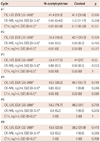
Another secondary endpoint of peak cardiac enzymes (CK, CK-MB and CTnI) at each cycle of chemotherapy is also similar in the two groups. All the cardiac enzymes did not increase through the whole chemotherapy schedules in both groups (Table 4).
No patients complained of symptoms related to arrhythmia like palpitation or escaped the beat during or after chemotherapy with ANT.

Clinical outcomes

There was no difference in terms of the composite all-cause mortality, HF and re-hospitalization at 1-, 3-, 6- and 12-month in the NAC and control groups (Fig. 2, Table 5).

Discussion

Risk factors for ANT-induced cardiomyopathy include the following; age greater than 70 years, diabetes, gender, hypertension, liver disease, poor nutrition, mediastinal radiotherapy, previous cardiac disease, or simultaneous administration of other antineoplastic agents, such as cyclophosphamides, actinomycin D, bleomycin, cisplatin, and methotrexate.4)5)13)14)
The most beneficial and convenient methods for preventing cardiomyopathy has been found with limitations on the amount of drugs used, and alternate drug-delivery methods, such as continuous infusion rather than rapid bolus injection. In a group of 630 patients receiving doxorubicin, Swain et al.15) found that HF developed in less than 5% of patients receiving a cumulative dose of 400 mg/m2 of body surface area, 16% of patients at a dose of 500 mg/m2, 26% of patients at a dose of 550 mg/m2, and 48% of patients at a dose of 700 mg/m2.13) An empirical dose limit of 500 mg/m2 of body surface area is suggested as a strategy to minimize the risk of ANT induced cardiomyopathy.16)
More actively, some potential antioxidative drugs have shown promise in animal models, although it is not clear how well these models predict human response. Those are sodium tanshinone IIA sulphonate, centella asiatica, vitamin E, vitamin A and probucol.5)9)17)18) Other agents, including erythropoietin,19) thrombopoietin20) and iloprost,21) also demonstrated positive results, although, they are small-sized animal studies.
In a clinical view point, enalapril and carvedilol appear promising.12)22) They may exert their effect through blocking oxidative stress caused by chemotherapy, as well as inhibiting renin-angiotensin-aldosterone systems or sympathetic nervous system, accompanied by hemodynamic change of the heart. However, the use of these drugs has some limitations to apply in clinical practice. They require a long time to get their efficacy as much as 6-12 months irrespective of status of having hypertension. As a consequence, compliance may be problematic, and there is no data regarding how the LV function can change after completion of the medications. More importantly, there is no supporting data thereafter. As such, it is yet too early to routinely recommend enalapril or carvedilol to protect the heart from cardiotoxicity.
N-acetylcysteine has been used for mucolytics and antidote for over dosed drugs in the emergency department, and some other medical area to derive antioxidative property because it has some merits for its very safe profile with low side effects, low cost and easy availability. Old data from an animal study with a mouse model demonstrated very promising results with short-term high dose NAC, especially just before chemotherapeutic agent administration. The survival rate was as high as 62.3% in NAC, as compared to 0% in the control group.9)
The dose of NAC use in this study is as high as 100 mg to 2000 mg/kg, and the efficacy is linearly increased in proportional to the dose used.9) However, there is no data of using NAC for preventing ANT-associated HF in humans. As far as we are aware, this is the first study to evaluate NAC, the representative antioxidant in ANT-induced cardiotoxicity. However, current prospective randomize trial does not demonstrate that NAC protects the heart form decrease of LVEF, and enlargement of LV and influence the mitral inflow pattern. On the contrary the NAC-treated group showed higher trend for an increase of LVESD, as compared to the control (p=0.064).
The rate of primary outcome occurrence is so little and this mainly attributed to the negative results of the whole study.
The first reason of negative results could be attributed to somewhat of a short period and small dose of NAC usage. We adopted the schedule and doses of NAC as 1200 mg 3-times a day for 2 days referring to other similar trials for the prevention of kidney function deterioration after intra-arterial administration of contrast media with NAC.23) Because the main mechanism of developing renal damage, by contrast media, is suggested as reactive oxygen free radicals, which are also the main plausible cause of ANT-induced cardiac toxicity. When being used in drug intoxication, the NAC are given even higher dose to neutralize toxicity; 150 mg/kg for full drip and 50 mg/kg for 4 hours, followed by 100 mg/kg for 16 hours, and totally 300 mg/kg for 20 hours. According to this regimen, the total dose of NAC for adult with body weight of 60 kg is 18000 mg. Moreover, considering the total dose of 2000 mg/kg in a mouse model in a positive animal study, our drug dose may be small.9)
The duration may be of importance. As mentioned earlier, in the clinical trial with positive results, researchers used enalapril or carvedilol for 6- to 12-month after chemotherapy. Therefore, the period of NAC exposure in the current study may not be adequate to exert full antioxidative effect.
Secondly, the study patients in EPOCH trial are at-low risk for developing cardiomyopathy. The mean age is relatively young (approximately 50 years old), has little risk factors of HF and relatively low dose of ANT used.
EPOCH trial has some limitations. Firstly, this study is under powered in statistics. We referred to the prior study dealing with the efficacy of enalapril on the prevention of ANT-cardiomyopathy, in estimating the number of study patients as described above.12) The primary outcome of EF decrease ≥10% unit associated with under 50% was the same criteria as our study. It occurred at a rate of 43% in the reference study.12) We assumed the primary outcome rate as 35%, considering it would occur less than that of the reference study, in which the sicker patients with elevated TnI after first chemotherapy were enrolled.12)
However, the incidence of ANT-induced cardiomyopathy in our study is too little, as compared to the pre-defined assumption. Consequently, our study is underpowered and the results are inconclusive in proving the NAC's efficacy.
Secondly, we did not measure the B-type nitriuretic peptide (BNP). Although echocardiography is the gold standard for detecting ANT-induced cardiomyopathy, some researchers suggest early detection may be possible by measuring the biomarkers like cTnI and CK-MB. We routinely measured the cTnI and CK-MB in each cycle, but not the BNP, mainly due to the limitations of research grants. Natriuretic peptides have shown promising results in the assessment and monitoring of acute, late clinical and subclinical damage of the myocardium, in association with chemotherapy. Thus, measuring BNP would have been beneficial in proving NAC's efficacy.24-26)
Thirdly, if we have measured and used tissue velocity and strain imaging, they also would have revealed benefit of NAC by early detection of preclinical change of LV systolic function before echocardiographic LVEF change.27)
Although the current study has failed in proving the efficacy of NAC, this makes us perceive that positive findings in-vivo and in-vitro study cannot always be translated into clinical settings. Future studies with high dose and/or longer duration regimen of NAC may resolve the query regarding the true effectiveness of NAC in ANT-associated cardiomyopathy.
In conclusion, this prospective randomized controlled trial cannot demonstrate that NAC prevents ANT induced cardiotoxicity. Although this study is underpowered, which is mainly attributed to the small primary event rate and could not prove the true efficacy of NAC, the use of NAC should be postponed until more supporting evidence is revealed.

Figures and Tables

Fig. 1
Rate of anthracycline induced cardiac toxicity define as 10% or more decrease of LVEF and concomitant EF <50% at 6-month between NAC and control group. LVEF: left ventricular ejection fraction, NAC: N-acetylcysteine.
kcj-43-174-g001
Fig. 2
Cumulative event rate at 12-month between NAC and control group. NAC: N-acetylcysteine.
kcj-43-174-g002
Table 1
Baseline characteristics of patients
kcj-43-174-i001

*Values are mean±standard deviation, Defined as systolic pressure ≥140 mm Hg and/or diastolic pressure ≥90 mm Hg or patients who have taken antihypertensive medications, Defined as total cholesterol level ≥240 mg/dL or patients who have taken statins. BMI: body mass index, LVEF: left ventricular ejection fraction, CAF: cyclophosphamide+adriamycine+fluorouracil, AC: adriamycin+cyclophosphamide, CEF: cyclophosphamide+epirubicine+fluorouracil, CHOP: cyclophosphamide+adriamycine+vincristine+presnisone, NAC: N-acetylcysteine, BUN: blood urea nitrogen, hs-CRP: high sensitivity C-reactive protein

Table 2
Changes in EF, LVESD and LVEDD following administration of N-acetylcysteine or control
kcj-43-174-i002

*Values are mean±standard deviation, The sum of the Ns (n=99) does not equal the total number of patients (n=103) in the intention-to-treat analysis because echocardiography was not performed in 4 patients. LVESD: left ventricular end systolic dimension, LVEDD: left ventricular end diastolic dimension, EF: ejection fraction

Table 3
Information on patients with anthracycline-induced cardiomyopathy
kcj-43-174-i003

BMI: body mass index, LVEF: left ventricular ejection fraction, hs-CRP: high sensitivity C-reactive protein, CAF: cyclophosphamide+adriamycine+fluorouracil, AC: adriamycin+cyclophosphamide, CEF: cyclophosphamide+epirubicine+fluorouracil

Table 4
Peak CK, CK-MB and CTnI at the end of each cycle of chemotherapy between N-acetylcysteine and control
kcj-43-174-i004

*The numbers in square brackets denote reference values. SD: standard deviation, CK-MB: creatine kinase-MB, cTnI: cardiac Troponin I

Table 5
Cumulative rate of clinical outcomes including all cause mortality, heart failure, stroke and heart failure hospitalization during admission, at 1-, 3, 6, and 12-month after completion of chemotherapy
kcj-43-174-i005

n/N: event number/total number

Acknowledgments

This study was supported by grants from the Korean Society of Cardiology (2008).

Notes

The authors have no financial conflicts of interest.

References

1. Hequet O, Le QH, Moullet I, et al. Subclinical late cardiomyopathy after doxorubicin therapy for lymphoma in adults. J Clin Oncol. 2004. 22:1864–1871.
2. Cardinale D, Sandri MT, Colombo A, et al. Prognostic value of troponin I in cardiac risk stratification of cancer patients undergoing high-dose chemotherapy. Circulation. 2004. 109:2749–2754.
3. Bristow MR, Billingham ME, Mason JW, Daniels JR. Clinical spectrum of anthracycline antibiotic cardiotoxicity. Cancer Treat Rep. 1978. 62:873–879.
4. Zucchi R, Danesi R. Cardiac toxicity of antineoplastic anthracyclines. Curr Med Chem Anticancer Agents. 2003. 3:151–171.
5. Takemura G, Fujiwara H. Doxorubicin-induced cardiomyopathy from the cardiotoxic mechanisms to management. Prog Cardiovasc Dis. 2007. 49:330–352.
6. Yeh ET, Tong AT, Lenihan DJ, et al. Cardiovascular complications of cancer therapy: diagnosis, pathogenesis, and management. Circulation. 2004. 109:3122–3131.
7. Deng S, Kruger A, Kleschyov AL, Kalinowski L, Daiber A, Wojnowski L. Gp91phox-containing NAD(P)H oxidase increases superoxide formation by doxorubicin and NADPH. Free Radic Biol Med. 2007. 42:466–473.
8. Spallarossa P, Altieri P, Garibaldi S, et al. Matrix metalloproteinase-2 and -9 are induced differently by doxorubicin in H9c2 cells: The role of MAP kinases and NAD(P)H oxidase. Cardiovasc Res. 2006. 69:736–745.
9. Doroshow JH, Locker GY, Ifrim I, Myers CE. Prevention of doxorubicin cardiac toxicity in the mouse by N-acetylcysteine. J Clin Invest. 1981. 68:1053–1064.
10. Rosenthal DS, Braunwald E. Braunwald E, editor. Cardiac effects of radiation therapy and chemotherapy. Heart Disease: A Textbook of Cardiovascular Medicine. 2001. Philadelphia, PA: WB Saunders Co.;1746–1751.
11. Pai VB, Nahata MC. Cardiotoxicity of chemotherapeutic agents: incidence, treatment and prevention. Drug Saf. 2000. 22:263–302.
12. Cardinale D, Colombo A, Sandri MT, et al. Prevention of high-dose chemotherapy-induced cardiotoxicity in high-risk patients by angiotensin-converting enzyme inhibition. Circulation. 2006. 114:2474–2481.
13. Lipshultz SE, Lipsitz SR, Mone SM, et al. Female sex and drug dose as risk factors for late cardiotoxic effects of doxorubicin therapy for childhood cancer. N Engl J Med. 1995. 332:1738–1743.
14. Steinherz LJ, Steinherz PG, Tan CT, Heller G, Murphy ML. Cardiac toxicity 4 to 20 years after completing anthracycline therapy. JAMA. 1991. 266:1672–1677.
15. Swain SM, Whaley FS, Ewer MS. Congestive heart failure in patients treated with doxorubicin: a retrospective analysis of three trials. Cancer. 2003. 97:2869–2879.
16. Figueredo VM. Chemical cardiomyopathies: the negative effects of medications and nonprescribed drugs on the heart. Am J Med. 2011. 124:480–488.
17. Hong HJ, Liu JC, Chen PY, Chen JJ, Chan P, Cheng TH. Tanshinone IIA prevents doxorubicin-induced cardiomyocyte apoptosis through Akt-dependent pathway. Int J Cardiol. 2012. 157:174–179.
18. Chao HH, Liu JC, Hong HJ, Lin JW, Chen CH, Cheng TH. L-carnitine reduces doxorubicin-induced apoptosis through a prostacyclin-mediated pathway in neonatal rat cardiomyocytes. Int J Cardiol. 2011. 146:145–152.
19. Li L, Takemura G, Li Y, et al. Preventive effect of erythropoietin on cardiac dysfunction in doxorubicin-induced cardiomyopathy. Circulation. 2006. 113:535–543.
20. Li K, Sung RY, Huang WZ, et al. Thrombopoietin protects against in vitro and in vivo cardiotoxicity induced by doxorubicin. Circulation. 2006. 113:2211–2220.
21. Neilan TG, Jassal DS, Scully MF, et al. Iloprost attenuates doxorubicin-induced cardiac injury in a murine model without compromising tumour suppression. Eur Heart J. 2006. 27:1251–1256.
22. Kalay N, Basar E, Ozdogru I, et al. Protective effects of carvedilol against anthracycline-induced cardiomyopathy. J Am Coll Cardiol. 2006. 48:2258–2262.
23. Tepel M, van der Giet M, Schwarzfeld C, Laufer U, Liermann D, Zidek W. Prevention of radiographic-contrast-agent-induced reductions in renal function by acetylcysteine. N Engl J Med. 2000. 343:180–184.
24. Feola M, Garrone O, Occelli M, et al. Cardiotoxicity after anthracycline chemotherapy in breast carcinoma: effects on left ventricular ejection fraction, troponin I and brain natriuretic peptide. Int J Cardiol. 2011. 148:194–198.
25. Lee CY, Burnett JC Jr. Natriuretic peptides and therapeutic applications. Heart Fail Rev. 2007. 12:131–142.
26. Urbanova D, Urban L, Danova K, Simkova I. Natriuretic peptides: biochemical markers of anthracycline cardiac toxicity? Oncol Res. 2008. 17:51–58.
27. Fallah-Rad N, Walker JR, Wassef A, et al. The utility of cardiac biomarkers, tissue velocity and strain imaging, and cardiac magnetic resonance imaging in predicting early left ventricular dysfunction in patients with human epidermal growth factor receptor II-positive breast cancer treated with adjuvant trastuzumab therapy. J Am Coll Cardiol. 2011. 57:2263–2270.
TOOLS
Similar articles