

 PDF
PDF ePub
ePub Citation
 Print
Print


Introduction
Valsalva aneurysms often remain undiagnosed until they rupture, usually into the right ventricle or atrium.1) The rupture may be clinically silent or present with symptoms of dyspnea and chest pain or with a new continuous to-and fro murmur. Transthoracic echocardiography (TTE) usually clinches the diagnosis, and a transesophageal echocardiography (TEE) or cardiac magnetic resonance imaging (MRI) may occasionally be necessary. On the other hand, a Valsalva aneurysm filled with thrombi can be difficult to accurately diagnose, because it mimics a cardiac tumor. We describe the rare case of a patient with a thrombosed ruptured Valsalva aneurysm who could be diagnosed preoperatively using both TEE and cardiac MRI.
Case
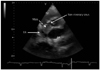
A 59-year-old man was referred to our hospital due to an abnormal electrocardiogram, atrial fibrillation and a complete atrioventricular blockage. He didn't feel any symptoms such as dyspnea on effort, chest pain or fatigue. He showed moderate cardiomegaly in a chest radiograph and non-significant findings in blood tests, including tumor markers. TTE showed an enlarged left ventricular dimension, deteriorating systolic function (ejection fraction=32%) and low echoic mass (26×30 mm) between the atrial septum and the non-coronary sinus (Fig. 1). A coronary angiography showed disease in three vessels, including chronic total occlusion of the proximal left anterior descending artery. No feeder artery to the cardiac mass was observed. Initially we planned to perform a biopsy of the mass using fluoroscopic and transesophageal echo-guidance, because we suspected the mass was a cardiac tumor.2) However, the patient refused the biopsy due to the high risk and invasiveness of the procedure. Thus, we performed noninvasive modalities, including cardiac MRI and TEE, in an attempt to make an accurate diagnosis of the mass.
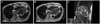
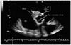
A cardiac MRI showed that the mass lesion presented high signal intensity mostly, mixed with a low focal signal intensity on a T2-weighted fast spin-echo image and a dark signal intensity on a T1-weighted fast spin-echo image (Fig. 2A and B). These MRI findings suggested that the mass was a subacute hemorrhage. In addition, the sagittal oblique cine MRI revealed that the mass was connected to the non-coronary sinus and was not located in the right atrium (Fig. 2C). A TEE showed a dilated non-coronary sinus filled with an irregular-surfaced low echoic mass (Fig. 3). By integration of the MRI findings of tissue characterization together with the accurate location of the mass with TEE findings of the irregular surface of the mass and a partial defect of the edge of the non-coronary sinus, we diagnosed that the cardiac mass was a thrombosed Valsalva aneurysm that had perforated the inter-atrial septum before. Surgery was performed.
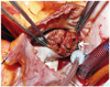
A non-coronary sinus plasty and a coronary-aorta bypass graft were performed. The operative findings revealed a defected and thrombi filled non-coronary sinus (Fig. 4), which coincided with the preoperative diagnosis from both the MRI and TEE. In addition, the patient underwent cardiac resynchronization therapy to address the complete atrioventricular block and reduced left ventricular function. The patient's postoperative clinical course was uneventful.
Discussion
There have been several reports of Valsalva aneurysms that could be diagnosed by the onset of atrioventricular blockage like what was seen in our case.3)4) Valsalva aneurysms have been reported to have other complications, including rupture,5) aortic regurgitation,6) outflow tract occulusion,7) myocardial ischemia8) and embolism.9) In our case, the site of the aneurysm was the non-coronary sinus, and it is usual that a non-coronary sinus aneurysm ruptures into the right atrium.10) Fortunately for this patient, the aneurysm perforation developed in only the non-coronary sinus and stopped at the right atrium wall. As a result, the patient had penetration into the inter-atrial septum.
A surgical repair is usually performed for both ruptured and unruptured Valsalva aneurysms. A Valsalva aneurysm filled with thrombi can be difficult to diagnose because it mimics a cardiac tumor.10) In this case, we were able to make a correct diagnosis for two reasons. First, the cardiac MRI showed that the mass was connected to the non-coronary sinus and was not located in the right atrium. The MRI has inferior spatial resolution (1.0-2.0 mm) as compared with a CT and echocardiography, but it has superior soft tissue contrast.11) The MRI findings of high signal intensity on a T2-weighted fast spin-echo image and dark signal intensity on a T1-weighted fast spin-echo image suggested that the mass was a subacute hemorrhage. Second, the TEE showed a defected non-coronary sinus, although an MRI could not show the defect accurately. In addition, the TEE showed that the surface of the mass in the non-coronary sinus was irregular, which we suspected indicated the presence of thrombi. The TEE was useful for real-time evaluation and spatial resolution (5-10 MHz). In conclusion, although it is usually difficult to make a diagnosis prior to the operation in these cases, both MRI and TEE findings were very useful for the preoperative diagnosis of the thrombosed ruptured Valsalva aneurysm mimicking a cardiac tumor.
 XML Download