Journal List > Korean Circ J > v.42(2) > 1016944

Kim and Ahn: A Long Road for Stem Cells to Cure Sick Hearts: Update on Recent Clinical Trials

Abstract

The contribution of stem cells to cure damaged hearts has finally been unraveled. A large number of preclinical and clinical studies have showed beneficial outcomes after myocardial infarction. In this review, the current understanding of stem cell therapy in preclinical and clinical experiences is summarized. Stem cells from bone marrow have shown a potential to improve cardiac performance after myocardial infarction in animal and early clinical studies. Clinical trials from all over the world have provided safety assessments of stem cell therapy with marginal improvement of clinical outcomes. Thus, further investigations should be encouraged to resolve the discrepancies between studies, clinical issues, and unclear translational findings. This review provides information and commentary on key trials for stem cell-based treat-ment of cardiovascular disease.

Introduction

According to a World Health Organization (WHO) report, cardiovascular diseases (CVDs) are the number one cause of death globally. It was estimated that 17.3 million people died from CVDs in 2008, representing 30% of all global deaths. In addition, it is predicted that almost 23.6 million people will die from CVDs by 2030 (WHO, 2011). The most important risk factors of CVDs are unhealthy diet, physical inactivity, smoking, and harmful use of alcohol. The effects of unhealthy diet and physical inactivity may show up in individuals as raised blood pressure, raised blood glucose, raised blood lipids, and overweight and obesity. These are called metabolic risk factors.
Rapid reperfusion of the culprit vessel for salvaging ischemic myocardium and optimal medications reduce complications and improve survival rate. Although many drugs and medical devices have been developed, the incidence of CVDs remains high. More efficacious therapeutic modalities need to be explored by medical researchers. Since the discovery of stem cells, a significant amount of research and development has emerged to be clinically applied for various incurable diseases including CVDs.
The human heart is a dynamic organ. Traditionally, the myocardium has been considered as terminally differentiated; however, there is growing evidence that cardiomyocytes are able to become proliferative when they face substantial damage such as myocardial infarction or heart failure.1)2) In addition to intrinsic repair mechanisms, progenitor/stem cell plasticity has emerged as one of the ways to be regenerated.3)
Stem cells are undifferentiated pluripotent multilineage cells with the ability to renew themselves. The sources of stem cells include the embryo, fetus, and various parts of adult tissues. Stem cells or progenitor cells are classified according to their characteristics in Table 1.
Many clinical trials showed excellent safety and feasibility of adult stem cells, but the efficacy of stem cell therapy is not satisfactory to improve cardiac function substantially.
The basic mechanisms of stem cell action on the injured myocardium will not be discussed here. We will focus instead on the current status of preclinical/clinical studies regarding therapeutic opportunities.
In this review, we summarize recent results of preclinical/clinical trials that have evaluated the safety, feasibility, and efficacy of cell therapy in heart disease.

Walking With Animal Study

Direct or indirect transplantation of adult stem cells to damaged hearts is emerging as an innovative strategy to ameliorate cardiac remodeling and dysfunction after acute myocardial infarction (AMI).
Potential sources of functional cardiomyocytes has been explored and utilized for cell therapy to replace injured cardiomyocytes. Stem cells are characterized by their ability to self-renew, clone, and differentiate into multiple tissues. Adult stem cells are isolated and characterized from various sources; peripheral blood,4) bone marrow (BM),5) adipose tissue,6) umbilical cord blood,7) amniotic membrane,8) and dental pulp.9)
A large number of animal studies have demonstrated that stem cells could be engrafted and differentiated within the heart.3)10-12) In preclinical studies, several large-animal species, including swine, sheep, and dogs, have been used to investigate the effects of stem cell therapy in CVDs models. Stem cells can be delivered to the heart by intravenous infusion, direct surgical injection, or catheter-based intracoronary infusion.
Orlic and colleagues first reported the repair of infarct myocardium through transplantation of bone marrow cells (BMCs) in mice.3)
Progenitors or stem cells with cardiomyogenic potential were studied both in vitro and in vivo. Experimental data showed the expression of cardiac markers such as cardiac contractile proteins in stem cells in transplanted myocardium. However, they were rarely found.
One of the popular strategies to increase the cardiomyogenesis of stem cells is the stimulation of stem cells with an anticancer drug, 5-azacytidine, which is a nonspecific deoxyribonucleic acid methylation inhibitor. Transient stimulation with 5-azacyticine for one day substantially increased cardiac protein expression in BM-derived mesenchymal stem cells (MSCs) and sca-1-positive adult cardiac stem cells with spontaneous beating on culture system.13)
To prove functional stem cell therapy, a small animal model has widely been used. Myocardial infarction was experimentally induced by surgical ligation of the coronary artery such as left anterior descending artery in mice or rats, sometimes with reperfusion. Various stem cell types, transplantation route, cell number, and transplantation timing have been studied in these small animal models. Many studies are reported that stem cell therapy is safe, feasible, and promising for the cure of MI. Histological data has revealed that the injected stem cells can survive in ischemic myocardium that participated in neovascularization and cardiomyogenesis. Scar size, cardiac remodeling, fibrosis, and inflammation were much more effectively improved. Cardiac function was also substantially improved after stem cell therapy. Based on these fantastic results of animal studies, many clinical studies were designed and initiated to transfer stem cell therapy to the bedside all over the world.
Various stem cells or progenitor cells have been introduced for cardiac repair in the last few years, although many past and ongoing clinical trials use predominantly adult autologous BM-derived cells. The use of BMCs in CVDs has the advantage that BM can be easily accessed, and isolated cells can be expanded for autologous application.
Experimental studies of cell priming revealed that it improved cell survival, retention, integration, and differentiation.12)14)15) In addition, genetic modification of stem cells before application with the prosurvival gene Akt,16) vascular endothelial growth factor,17) or fibroblast growth factor 218) promoted therapeutic efficacy.
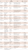
Table 2 shows summarized results of MSCs therapy in porcine AMI model. The anatomy and physiology of the porcine heart is well known to be similar to a human heart, and it is considered that the porcine MI model is the best model for CVDs research. Cell number, delivery procedure, and surgical techniques in the porcine model are also similar to the clinical setting, and more realistic implications could be provided compared with a small animal model.

Running With Clinical Study

In AMI, cardiac muscle is damaged to become dysfunctional. After successful percutaneous coronary intervention (PCI) and coronary artery bypass graft surgery with optimal medications, cardiac function is restored only to a limited degree {3-4% improvement in left ventricular ejection fraction (LVEF)} which may result in cardiac remodeling in approximately 60% of the patients with myocardial infarction.19)20)
Many clinical studies have proceeded for proving their safety and efficacy to reach a final goal "new therapy". Regarding cell type, most clinical trials have used unfractionated BMCs as the delivery product, postulating that stem/progenitor cells are the biologically applicable therapeutic agents. The most widely applicable technique for stem cell delivery is intracoronary infusion from a clinical standpoint.
The first human clinical trial of stem cell trial was intracoronary infusion of autologous BM unfractionated mononuclear cells to AMI patient.21) Subsequent clinical studies of stem cells for AMI were then initiated.
A variety of studies have demonstrated significant improvement of ventricular performance after stem cell therapy in AMI, resulting in an increase in LVEF and decrease in infarct size. In most cases, stem cell transplantation was performed in a time frame of 12 hours to several days after MI. Although there is large variability of hemodynamic data after cell therapy, there is moderate improvement of cardiac performance by stem cell therapy that is more quantitatively effective than therapeutic interventions and pharmacotherapy.22) Thus, autologous stem cell therapy represents an innovative and effective procedure for regeneration of impaired hearts in the early phase after the infarct.
As seen in Table 3, most recent clinical trials utilized BM-derived mononuclear cells isolated from patients after PCI. BM has been considered the safest source for autologous transplantation of stem cells, usually mononuclear cells, in clinical trials. For now the most widely used clinically approved source for stem cell therapy is autologous stem cells from BM (www.clinicaltrials.gov).
In addition to BM-mononuclear cells, MSCs are now actively under investigation for cardiac repair. BM contains a population of hematopoietic stem cells and a rare population of plastic-adherent stromal cells (1 in 10000 nucleated cells in BM).23)
These plastic adherent cells are MSCs capable of forming singlecell colonies, expansion in culture, differentiation into osteoblasts, chondrocytes, and adipocytes.24)25) MSCs were shown to be differentiated into a myogenic phenotype.13) Animal studies demonstrated that human MSCs could be transdifferentiated into endoderm-derived cells10) in injected myocardium, and coculture of MSC with ventricular myocytes induced transdifferentiation into a cardiomyocyte phenotype in vitro.26) Large-animal preclinical studies of MSCs administration in post-MI heart demonstrated the ability of MSCs to engraft, differentiate, and produce substantial functional recovery.12)16)27)28)
The therapeutic effect of MSC on AMI has been reported in four clinical trials. Chen et al.29) infused autologous MSC by intracoronary route and demonstrated regional wall motion and global LVEF were improved after six months of cell therapy. At that time, Vulliet et al.30) reported that a microinfarction occurred after intracoronary infusion of MSCs in a dog MI model. A Prochymal trial31) was designed to evaluate the safety of intravenous application of allogeneic BM-derived MSCs to AMI patients. According to animal studies, a large proportion of infused cells were trapped in the lungs after administration, raising potential concerns regarding compromised pulmonary function.32) The results of the Prochymal trial did not show any evidence of a pulmonary safety risk after infusion of allogeneic human (h)BM-MSCs. Instead, those data revealed improved pulmonary function in the MSC-treated patients, compared with baseline status. The rate of arrhythmia event was 4-fold lower in the hMSCs group than in the placebo group (8.8% vs. 36.8%, p=0.025). In ischemic cardiomyopathy, transendocardial injection of autologous BM-derived progenitor cells including mononuclear cells or MSCs produced functional recovery in scarred myocardium and reversed remodeling of the LV chamber.33) Unfortunately, however, they did not determine superiority between mononuclear cells and MSCs. Without the placebo control group, data from only 4 patients in each group were analyzed. The transendocardial autologous cells in an ischemic heart failure trial (TAC-HFT) and Percutaneous Stem Cell Injection Delivery Effects on Neomyogenesis (POSEIDON) trials34) are in progress to intensify those limitations (www.clinicaltrials.gov). TAC-HFT is designed for comparing BM-MSCs versus mononuclear cells, and POSEIDON is comparing the effects of allogeneic versus autologous MSC therapy in ischemic cardiomyopathy patients.
The delivery of MultiStem, an allogeneic BM-derived stem cell produced by Athersys, Inc. (Cleveland, OH, USA), to AMI patients has proved to be safe and well tolerated.35) They showed MultiStem delivered via transarterial adventitia using a microsyringe catheter was safe with improvement of cardiac function in a dose-dependent manner.
Although the exact mechanism of MSC effects is still unresolved, accumulating results of large animal studies and clinical studies have shown that MSC-based therapy for cardiac repair is safe and provides substantial improvement in cardiac structure and function.

Looking Back With Consideration

The therapeutic effect of stem cell therapy on heart disease has been shown by experimental studies using small animal models. Results have been reported as extraordinarily promising with experimental results such as cardiomyogenesis, neovascularization, and paracrine effect on injured myocardium. Now we are facing challenges for bringing stem cells to clinically applicable therapeutics. Intracoronary transfer of autologous BMCs after optimum reperfusion therapy does not dramatically augment recovery of global LV function in patients, but could favorably affect cardiac remodeling after MI. Mixed results have been reported in clinical trials of stem cell administered patients after AMI with minimal improvement of ejection fraction or only a transient clinical benefit.
The different outcomes were attributed to differences in cell preparations, timing and method of cell administration, choice of endpoints, and characteristics of patients. Some studies failed to determine persistent clinical benefits after stem cell application (Table 3).
Arrhythmias have been reported to be associated with intramyocardial rather than intracoronary injection of stem cells in the early clinical studies; intramyocardial injection could be responsible for arrhythmogenesis. In addition, local injection induces a highly uneven distribution of cells, at least early after injection, which increases electrophysiological heterogeneity. Although recent available results of clinical experience35)36) so far suggest that proarrhythmic effects may be transient, cardiac arrhythmia occurs unpredictably, and long-term follow-up studies would be essential to understand the arrhythmogenesis induced by stem cell transplantation.
The most effective and safe cell type for myocardial repair and the clinical significance of cell therapy-induced arrhythmias will be determined in future pre-clinical and clinical studies.
There was a case report about fatal events after autologous hematopoietic stem cell application in a lupus nephritis patient.37) After direct renal injection of stem cells isolated from peripheral blood, masses at the sites of injection were developed with hematuria. Pathologic analysis revealed the masses were angiomyeloproliferative lesions and suggested to be a possible complication of stem cell therapy. There was no way to find out the detailed cause of death in those cases, but stem cell transplantation could be a causative event.
The ultimate goal of cell therapy is the regeneration of lost cardiac muscle along with the reversal of adverse remodeling. Despite growing clinical experience, the absence of standardized clinical end point in human trials has left us with fundamental questions. Issues to be addressed in the future include determining the ideal cell type, the cell number to be delivered, optimal cell isolation method, efficient cell storage, and optimal time of administration to improve the efficacy of the therapy. After that, more realistic and optimized conditions of stem cell therapy will be applied to patients suffered from CVDs with guaranteed safety.
The risk of exposing patients to possible adverse outcomes of cell therapy must be seriously considered before clinical application. The argument that clinical trials should be delayed till mechanisms are perfectly understood will deprive a large number of patients from therapeutic chances that may bring them clinical recovery. Stem cell therapy is a novel and innovative approach to cardiac therapeutics which has been achieved by numerous preclinical and early clinical studies showing safety, feasibility, and early efficacy.

Conclusion

Recent evidence from studies in animals and humans demonstrates the important roles of stem cells in CVDs. In this review, reports of recent clinical trials of stem cell therapy for myocardial infarction are summarized and some important considerations are suggested for further application. For now, the challenge is to improveme the scientific concept to clinical setting with current treatment modalities.

Figures and Tables

Table 1
Major cell types with potential for cardiac cell therapy
kcj-42-71-i001

Marin-Garcia M. Heart failure: bench to bedside. New York: Springer; 2010

Table 2
Summary of results from mesenchymal stem cell therapy cases in the swine model
kcj-42-71-i002

BM: bone marrow, CMR: cardiac magnetic resonance imaging, EDV: end-diastolic volume, EC: endocardial injection, EF: ejection fraction, EPC: endothelial progenitor cells, HGF: hepatocyte growth factor, IC: intracoronary infusion, IGF-1: insulin like growth factor-1, IM: intramyocardial injection, LAD: left anterior descending artery, MNC: mononuclear cells, MPC: multipotent progenitor cells, MSC: mesenchymal stem cells, PBMNC: peripheral blood-derived mononuclear cells, PET-CT: positron emission tomography-computed tomography, SPECT: single-photon emission computed tomography, MI: myocardial infarction, IV: intravenous, FISH: fluorescence in situ hybridization, F/U: follow-up

Table 3
Summary of recent stem cell therapy trials in myocardial infarction (and heart failure)
kcj-42-71-i003

BM: bone marrow, EDV: end-diastolic volume, EF: ejection fraction, IC: intracoronary infusion, MNC: mononuclear cells, MSC: mesenchymal stem cells, BMC: bone marrow cell, MI: myocardial infarction, PCI: percutaneous coronary intervention, STEMI: ST segment elevation myocardial infarction, HF: heart failure, DES: drug-eluting stent, LVEF: left ventricular ejection fraction, LVEDV: left ventricular end diastolic volume, LVESV: left ventricular end systolic volume, ESV: end-systolic volume, AMI: acute myocardial infarction, CABG: coronary artery bypass graft, PB: peripheral blood, LV: left ventricle, G-CSF: granulocyte colony stimulating factor, PBSC: peripheral blood stem cell, LVEDP: left ventricular end diastolic pressure, CSC: cardiac stem cell

Acknowledgments

This work was supported by a grant of the National Research Foundation of Korea funded by the Korean Government (MEST), Republic of Korea (2010-0020261), and by a grant of the Korea Healthcare Technology R&D Project, Ministry for Health, Welfare & Family Affairs, Republic of Korea (A084869).

Notes

The authors have no financial conflicts of interest.

References

1. Kajstura J, Leri A, Finato N, Di Loreto C, Beltrami CA, Anversa P. Myocyte proliferation in end-stage cardiac failure in humans. Proc Natl Acad Sci U S A. 1998. 95:8801–8805.
2. Beltrami AP, Urbanek K, Kajstura J, et al. Evidence that human cardiac myocytes divide after myocardial infarction. N Engl J Med. 2001. 344:1750–1757.
3. Orlic D, Kajstura J, Chimenti S, et al. Bone marrow cells regenerate infarcted myocardium. Nature. 2001. 410:701–705.
4. Wojakowski W, Tendera M, Michałowska A, et al. Mobilization of CD34/CXCR4+, CD34/CD117+, c-met+ stem cells, and mononuclear cells expressing early cardiac, muscle, and endothelial markers into peripheral blood in patients with acute myocardial infarction. Circulation. 2004. 110:3213–3220.
5. Kucia M, Reca R, Jala VR, Dawn B, Ratajczak J, Ratajczak MZ. Bone marrow as a home of heterogenous populations of nonhematopoietic stem cells. Leukemia. 2005. 19:1118–1127.
6. Zuk PA, Zhu M, Ashjian P, et al. Human adipose tissue is a source of multipotent stem cells. Mol Biol Cell. 2002. 13:4279–4295.
7. Carlin R, Davis D, Weiss M, Schultz B, Troyer D. Expression of early transcription factors Oct-4, Sox-2 and Nanog by porcine umbilical cord (PUC) matrix cells. Reprod Biol Endocrinol. 2006. 4:8.
8. De Coppi P, Bartsch G Jr, Siddiqui MM, et al. Isolation of amniotic stem cell lines with potential for therapy. Nat Biotechnol. 2007. 25:100–106.
9. Kerkis I, Kerkis A, Dozortsev D, et al. Isolation and characterization of a population of immature dental pulp stem cells expressing OCT-4 and other embryonic stem cell markers. Cells Tissues Organs. 2006. 184:105–116.
10. Toma C, Pittenger MF, Cahill KS, Byrne BJ, Kessler PD. Human mesenchymal stem cells differentiate to a cardiomyocyte phenotype in the adult murine heart. Circulation. 2002. 105:93–98.
11. Silva GV, Litovsky S, Assad JA, et al. Mesenchymal stem cells differentiate into an endothelial phenotype, enhance vascular density, and improve heart function in a canine chronic ischemia model. Circulation. 2005. 111:150–156.
12. Kim YS, Kwon JS, Hong MH, et al. Promigratory activity of oxytocin on umbilical cord blood-derived mesenchymal stem cells. Artif Organs. 2010. 34:453–461.
13. Wakitani S, Saito T, Caplan AI. Myogenic cells derived from rat bone marrow mesenchymal stem cells exposed to 5-azacytidine. Muscle Nerve. 1995. 18:1417–1426.
14. Hahn JY, Cho HJ, Kang HJ, et al. Pre-treatment of mesenchymal stem cells with a combination of growth factors enhances gap junction formation, cytoprotective effect on cardiomyocytes, and therapeutic efficacy for myocardial infarction. J Am Coll Cardiol. 2008. 51:933–943.
15. Kim YS, Ahn Y, Kwon JS, et al. Priming of Mesenchymal Stem Cells with Oxytocin Enhances the Cardiac Repair in Ischemia/Reperfusion Injury. Cells Tissues Organs. 2012. in press.
16. Lim SY, Kim YS, Ahn Y, et al. The effects of mesenchymal stem cells transduced with Akt in a porcine myocardial infarction model. Cardiovasc Res. 2006. 70:530–542.
17. Deuse T, Peter C, Fedak PW, et al. Hepatocyte growth factor or vascular endothelial growth factor gene transfer maximizes mesenchymal stem cell-based myocardial salvage after acute myocardial infarction. Circulation. 2009. 120:11 Suppl. S247–S254.
18. Song H, Kwon K, Lim S, et al. Transfection of mesenchymal stem cells with the FGF-2 gene improves their survival under hypoxic conditions. Mol Cells. 2005. 19:402–407.
19. Abbate A, Biondi-Zoccai GG, Appleton DL, et al. Survival and cardiac remodeling benefits in patients undergoing late percutaneous coronary intervention of the infarct-related artery: evidence from a meta-analysis of randomized controlled trials. J Am Coll Cardiol. 2008. 51:956–964.
20. Konstam MA, Kramer DG, Patel AR, Maron MS, Udelson JE. Left ventricular remodeling in heart failure: current concepts in clinical significance and assessment. JACC Cardiovasc Imaging. 2011. 4:98–108.
21. Strauer BE, Brehm M, Zeus T, et al. Intracoronary, human autologous stem cell transplantation for myocardial regeneration following myocardial infarction. Dtsch Med Wochenschr. 2001. 126:932–938.
22. Schächinger V, Erbs S, Elsässer A, et al. Improved clinical outcome after intracoronary administration of bone-marrow-derived progenitor cells in acute myocardial infarction: final 1-year results of the REPAIR-AMI trial. Eur Heart J. 2006. 27:2775–2783.
23. Friedenstein AJ, Chailakhjan RK, Lalykina KS. The development of fibroblast colonies in monolayer cultures of guinea-pig bone marrow and spleen cells. Cell Tissue Kinet. 1970. 3:393–403.
24. Friedenstein AJ, Chailakhyan RK, Latsinik NV, Panasyuk AF, Keiliss-Borok IV. Stromal cells responsible for transferring the microenvironment of the hemopoietic tissues: cloning in vitro and retransplantation in vivo. Transplantation. 1974. 17:331–340.
25. Caplan AI. Molecular and cellular differentiation of muscle, cartilage, and bone in the developing limb. Prog Clin Biol Res. 1986. 217B:307–318.
26. Xu W, Zhang X, Qian H, et al. Mesenchymal stem cells from adult human bone marrow differentiate into a cardiomyocyte phenotype in vitro. Exp Biol Med (Maywood). 2004. 229:623–631.
27. Quevedo HC, Hatzistergos KE, Oskouei BN, et al. Allogeneic mesenchymal stem cells restore cardiac function in chronic ischemic cardiomyopathy via trilineage differentiating capacity. Proc Natl Acad Sci U S A. 2009. 106:14022–14027.
28. Wang D, Zhang F, Shen W, et al. Mesenchymal stem cell injection ameliorates the inducibility of ventricular arrhythmias after myocardial infarction in rats. Int J Cardiol. 2011. 152:314–320.
29. Chen SL, Fang WW, Ye F, et al. Effect on left ventricular function of intracoronary transplantation of autologous bone marrow mesenchymal stem cell in patients with acute myocardial infarction. Am J Cardiol. 2004. 94:92–95.
30. Vulliet PR, Greeley M, Halloran SM, MacDonald KA, Kittleson MD. Intra-coronary arterial injection of mesenchymal stromal cells and microinfarction in dogs. Lancet. 2004. 363:783–784.
31. Hare JM, Traverse JH, Henry TD, et al. A randomized, double-blind, placebo-controlled, dose-escalation study of intravenous adult human mesenchymal stem cells (prochymal) after acute myocardial infarction. J Am Coll Cardiol. 2009. 54:2277–2286.
32. Chin BB, Nakamoto Y, Bulte JW, Pittenger MF, Wahl R, Kraitchman DL. 111In oxine labelled mesenchymal stem cell SPECT after intravenous administration in myocardial infarction. Nucl Med Commun. 2003. 24:1149–1154.
33. Williams AR, Trachtenberg B, Velazquez DL, et al. Intramyocardial stem cell injection in patients with ischemic cardiomyopathy: functional recovery and reverse remodeling. Circ Res. 2011. 108:792–796.
34. Trachtenberg B, Velazquez DL, Williams AR, et al. Rationale and design of the Transendocardial Injection of Autologous Human Cells (bone marrow or mesenchymal) in Chronic Ischemic Left Ventricular Dysfunction and Heart Failure Secondary to Myocardial Infarction (TAC-HFT) trial: a randomized, double-blind, placebo-controlled study of safety and efficacy. Am Heart J. 2011. 161:487–493.
35. Perin EC, Dohmann HF, Borojevic R, et al. Transendocardial, autologous bone marrow cell transplantation for severe, chronic ischemic heart failure. Circulation. 2003. 107:2294–2302.
36. Stamm C, Westphal B, Kleine HD, et al. Autologous bone-marrow stem-cell transplantation for myocardial regeneration. Lancet. 2003. 361:45–46.
37. Thirabanjasak D, Tantiwongse K, Thorner PS. Angiomyeloproliferative lesions following autologous stem cell therapy. J Am Soc Nephrol. 2010. 21:1218–1222.
38. Shake JG, Gruber PJ, Baumgartner WA, et al. Mesenchymal stem cell implantation in a swine myocardial infarct model: engraftment and functional effects. Ann Thorac Surg. 2002. 73:1919–1925.
39. Amado LC, Saliaris AP, Schuleri KH, et al. Cardiac repair with intramyocardial injection of allogeneic mesenchymal stem cells after myocardial infarction. Proc Natl Acad Sci U S A. 2005. 102:11474–11479.
40. Price MJ, Chou CC, Frantzen M, et al. Intravenous mesenchymal stem cell therapy early after reperfused acute myocardial infarction improves left ventricular function and alters electrophysiologic properties. Int J Cardiol. 2006. 111:231–239.
41. Freyman T, Polin G, Osman H, et al. A quantitative, randomized study evaluating three methods of mesenchymal stem cell delivery following myocardial infarction. Eur Heart J. 2006. 27:1114–1122.
42. Feygin J, Mansoor A, Eckman P, Swingen C, Zhang J. Functional and bioenergetic modulations in the infarct border zone following autologous mesenchymal stem cell transplantation. Am J Physiol Heart Circ Physiol. 2007. 293:H1772–H1780.
43. Schuleri KH, Amado LC, Boyle AJ, et al. Early improvement in cardiac tissue perfusion due to mesenchymal stem cells. Am J Physiol Heart Circ Physiol. 2008. 294:H2002–H2011.
44. Hashemi SM, Ghods S, Kolodgie FD, et al. A placebo controlled, dose-ranging, safety study of allogenic mesenchymal stem cells injected by endomyocardial delivery after an acute myocardial infarction. Eur Heart J. 2008. 29:251–259.
45. Gyöngyösi M, Blanco J, Marian T, et al. Serial noninvasive in vivo positron emission tomographic tracking of percutaneously intramyocardially injected autologous porcine mesenchymal stem cells modified for transgene reporter gene expression. Circ Cardiovasc Imaging. 2008. 1:94–103.
46. Wolf D, Reinhard A, Seckinger A, Katus HA, Kuecherer H, Hansen A. Dose-dependent effects of intravenous allogeneic mesenchymal stem cells in the infarcted porcine heart. Stem Cells Dev. 2009. 18:321–329.
47. Moscoso I, Barallobre J, de Ilarduya OM, et al. Analysis of different routes of administration of heterologous 5-azacytidine-treated mesenchymal stem cells in a porcine model of myocardial infarction. Transplant Proc. 2009. 41:2273–2275.
48. Ly HQ, Hoshino K, Pomerantseva I, et al. In vivo myocardial distribution of multipotent progenitor cells following intracoronary delivery in a swine model of myocardial infarction. Eur Heart J. 2009. 30:2861–2868.
49. Schuleri KH, Feigenbaum GS, Centola M, et al. Autologous mesenchymal stem cells produce reverse remodelling in chronic ischaemic cardiomyopathy. Eur Heart J. 2009. 30:2722–2732.
50. Dubois C, Liu X, Claus P, et al. Differential effects of progenitor cell populations on left ventricular remodeling and myocardial neovascularization after myocardial infarction. J Am Coll Cardiol. 2010. 55:2232–2243.
51. Ellison GM, Torella D, Dellegrottaglie S, et al. Endogenous cardiac stem cell activation by insulin-like growth factor-1/hepatocyte growth factor intracoronary injection fosters survival and regeneration of the infarcted pig heart. J Am Coll Cardiol. 2011. 58:977–986.
52. van Ramshorst J, Bax JJ, Beeres SL, et al. Intramyocardial bone marrow cell injection for chronic myocardial ischemia: a randomized controlled trial. JAMA. 2009. 301:1997–2004.
53. Meyer GP, Wollert KC, Lotz J, et al. Intracoronary bone marrow cell transfer after myocardial infarction: 5-year follow-up from the randomized-controlled BOOST trial. Eur Heart J. 2009. 30:2978–2984.
54. Tendera M, Wojakowski W, Ruzyłło W, et al. Intracoronary infusion of bone marrow-derived selected CD34+CXCR4+ cells and non-selected mononuclear cells in patients with acute STEMI and reduced left ventricular ejection fraction: results of randomized, multicentre Myocardial Regeneration by Intracoronary Infusion of Selected Population of Stem Cells in Acute Myocardial Infarction (REGENT) Trial. Eur Heart J. 2009. 30:1313–1321.
55. Beitnes JO, Hopp E, Lunde K, et al. Long-term results after intracoronary injection of autologous mononuclear bone marrow cells in acute myocardial infarction: the ASTAMI randomised, controlled study. Heart. 2009. 95:1983–1989.
56. Assmus B, Rolf A, Erbs S, et al. Clinical outcome 2 years after intracoronary administration of bone marrow-derived progenitor cells in acute myocardial infarction. Circ Heart Fail. 2010. 3:89–96.
57. Grajek S, Popiel M, Gil L, et al. Influence of bone marrow stem cells on left ventricle perfusion and ejection fraction in patients with acute myocardial infarction of anterior wall: randomized clinical trial: impact of bone marrow stem cell intracoronary infusion on improvement of microcirculation. Eur Heart J. 2010. 31:691–702.
58. Arnold R, Villa A, Gutiérrez H, et al. Absence of accelerated atherosclerotic disease progression after intracoronary infusion of bone marrow derived mononuclear cells in patients with acute myocardial infarction: angiographic and intravascular ultrasound: results from the TErapia Celular Aplicada al Miocardio Pilot study. Am Heart J. 2010. 159:1154.e1–1154.e8.
59. Strauer BE, Yousef M, Schannwell CM. The acute and long-term effects of intracoronary Stem cell Transplantation in 191 patients with chronic heARt failure: the STAR-heart study. Eur J Heart Fail. 2010. 12:721–729.
60. Seth S, Bhargava B, Narang R, et al. The ABCD (Autologous Bone Marrow Cells in Dilated Cardiomyopathy) trial a long-term follow-up study. J Am Coll Cardiol. 2010. 55:1643–1644.
61. Traverse JH, McKenna DH, Harvey K, et al. Results of a phase 1, randomized, double-blind, Placebo-controlled trial of bone marrow mononuclear stem cell administration in patients following ST-elevation myocardial infarction. Am Heart J. 2010. 160:428–434.
62. Santoso T, Irawan C, Alwi I, et al. Safety and feasibility of combined granulocyte colony stimulating factor and erythropoetin based-stem cell therapy using intracoronary infusion of peripheral blood stem cells in patients with recent anterior myocardial infarction: one-year follow-up of a phase 1 study. Acta Med Indones. 2011. 43:112–121.
63. Mansour S, Roy DC, Bouchard V, et al. One-year safety analysis of the COMPARE-AMI Trial: comparison of intracoronary injection of CD133 bone marrow stem cells to Placebo in patients after acute myocardial infarction and left ventricular dysfunction. Bone Marrow Res. 2011. 2011:385124.
64. Hirsch A, Nijveldt R, van der Vleuten PA, et al. Intracoronary infusion of mononuclear cells from bone marrow or peripheral blood compared with standard therapy in patients after acute myocardial infarction treated by primary percutaneous coronary intervention: results of the randomized controlled HEBE trial. Eur Heart J. 2011. 32:1736–1747.
65. Penn MS, Ellis S, Gandhi S, et al. Adventitial delivery of an allogeneic bone marrow-derived adherent stem cell in acute myocardial infarction: phase I clinical study. Circ Res. 2012. 110:304–311.
66. Bolli R, Chugh AR, D'Amario D, et al. Cardiac stem cells in patients with ischaemic cardiomyopathy (SCIPIO): initial results of a randomised phase 1 trial. Lancet. 2011. 378:1847–1857.
67. Moreira Rde C, Haddad AF, Silva SA, et al. Intracoronary stem-cell injection after myocardial infarction: microcirculation sub-study. Arq Bras Cardiol. 2011. 97:420–426.
68. Solheim S, Seljeflot I, Lunde K, et al. The influence of intracoronary injection of bone marrow cells on prothrombotic markers in patients with acute myocardial infarction. Thromb Res. 2011. 12. 20. [Epub]. http://dx.doi.org/10.1016/j.thromres.2011.11.045.
69. Roncalli J, Mouquet F, Piot C, et al. Intracoronary autologous mononucleated bone marrow cell infusion for acute myocardial infarction: results of the randomized multicenter BONAMI trial. Eur Heart J. 2011. 32:1748–1757.
70. Ahmadi H, Moshkani Farahani M, Kouhkan A, et al. Five-Year Follow-up Of The Local Autologous Transplantation of CD133+ Enriched Bone Marrow Cells in Patients with Myocardial Infarction. Arch Iran Med. 2012. 15:32–35.
TOOLS
Similar articles