

 PDF
PDF ePub
ePub Citation
 Print
Print


Introduction
Left ventricular hypertrabeculation/noncompaction (LVHT) is characterized by a pattern of prominent trabecular meshwork and deep intertrabecular recesses that communicate with the left ventricular (LV) cavity. It is thought to be caused by arrest of normal embryogenesis of the endocardium and myocardium. Arrhythmias are common in patients with LVHT. Those arrhythmias frequently associated with LVHT include ventricular tachycardia (VT), atrial fibrillation (AF), atrioventricular (AV) block, QT prolongation, and Wolff-Parkinson-White (WPW) syndrome.1) In this report, we describe an unusual case of LVHT that was accompanied by severe mitral regurgitation, a fasciculo-ventricular accessory pathway, and atrial flutter (AFL). To the best of our knowledge, such a combination has not been previously reported. The following is a detailed report of the case along with a brief review of the relevant previous studies.
Case
A 29-year-old male visited our outpatient department due to exertional dyspnea for 3 weeks. He also complained of frequent and sustained episodes of palpitations associated with shortness of breath. He had no other medical history. The patient's blood pressure was 110/70 mm Hg, his pulse rate was 51 beats per minute, and his respiratory rate was 20 per minute. On physical examination, a systolic murmur of grade III/IV was audible at the apex. He had mild edema in the lower legs and the level of B-type natriuretic peptide was 276 pg/mL.
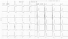
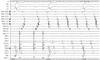
The chest posteroanterior showed cardiomegaly and mild pulmonary edema. An electrocardiography (ECG) demonstrated a sinus rhythm, pre-excitation and bizarre ST and T wave abnormality (Fig. 1). The positive delta indicating anteroseptal accessory pathway was discernable on the precordial leads.
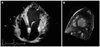
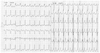
The apical 4-chamber view of a 2-dimensional echocardiogram presented LV hypertrophy and the LV ejection fraction was 36%. Prominent trabeculations and deep recesses were shown in the left ventricles and the recesses were perfused by intraventricular flow (Fig. 2A). The color Doppler echocardiogram showed severe mitral regurgitation. Later the cardiac magnetic resonance imaging was performed and it revealed sponge-shaped deep trabeculations in the anterior wall of the LV (Fig. 2B). At this portion, thickness of wall is about 8 mm and the trabeculation is 17 mm. Subendocardial delayed enhancement is noted in the LV. LVHT was diagnosed by Jenni et al.'s2) criteria. During the echocardiographic examination, tachycardia developed spontaneously and the ECG showed wide complex tachycardia with the QRS morphology, which was similar to the sinus rhythm (Fig. 3). The differential diagnosis included antidromic AV reentrant tachycardia, AFL, and atrial tachycardia. The patient underwent direct current cardioversion and was treated with angiotensin-converting enzyme inhibitor, diuretics, and digoxin.
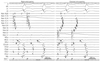
The patient underwent an electrophysiologic study. The electrode catheters were placed in the right atrium, in the coronary sinus, at the bundle of His, and in the right ventricular apex. The AH and HV intervals were 48 ms and 28 ms, respectively. Decremental atrial pacing caused prolongation of both AH and AV intervals in parallel. The HV interval and the degree of pre-excitation remained unchanged from both the right atrial and coronary sinus pacing with atrial extrastimuli (Fig. 4). The bundle of His pacing failed to normalize the QRS width. The diagnosis of fasciculo-ventricular accessory pathway was made. Decremental pacing on the proximal coronary sinus area induced typical AFL (Fig. 5), which was identical to the clinical tachycardia. A line of block was made across the cavo-tricuspid isthmus. Complete electrical blockade was confirmed by pacing both the low right atrium and proximal coronary sinus, resulting in late atrial activation on the opposite side.
The patient was discharged and is being regularly followed at the outpatient department. Screening the patient's parents and siblings demonstrated no structural heart disease. Warfarin was added to prevent thromboembolic events.
Discussion
Left ventricular hypertrabeculation/noncompaction is characterized by trabeculations and recesses within the ventricular myocardium, most commonly affecting the LV. The symptoms are mainly due to LV systolic dysfunction, arrhythmia, and thromboembolic complications.3) It is not clear whether LVHT is a distinct cardiomyopathy or a particular phenotypic expression of different cardiomyopathies.4)
Left ventricular hypertrabeculation/noncompaction can present in both children and adults. The most remarkable difference is the lack of facial dysmorphism in the adult population.5) A genetic study also showed some difference. An autosomal dominant trait was described in adults, while X-linked, autosomal dominant, and mitochondrial inheritance have been reported in children.6) Familial recurrence seems to be more common in adult patients than in children.
Stöllberger and Finsterer1) have analyzed the arrhythmias associated with LVHT. The prevalence differs between children and adults. In adults, the most frequent arrhythmias are VT, AF, QT prolongation and AV block. Ventricular pre-excitation was more frequently reported in children than in adults. Most of the ventricular pre-excitation was WPW syndrome. Mahaim fiber was reported in only one adult case. LVHT with AFL has only been reported in 4 patients. Three of them were adults. To our knowledge, this is the first case that presented with AFL and Mahaim-type accessory pathway as a bystander.
It is unknown whether AF/AFL shares a common pathogenesis with LVHT. However, one can assume that AF/AFL could be a secondary change of LV systolic or diastolic dysfunction, since this arrhythmia seldom occurs in childhood patients with LVHT. The present case was accompanied with severe mitral regurgitation. This might have additively affected the development of AFL. Although AFL was successfully ablated and the pre-excitation was proved to be a bystander, there is a possibility of developing AF in this patient. Thus, anticoagulation treatment was warranted to prevent thromboembolic events.
 XML Download