

 PDF
PDF ePub
ePub Citation
 Print
Print


Introduction
Left ventricular free wall rupture (LVFWR) is reported to occur in 2-6% of acute myocardial infarction (MI) cases. LVFWR is presumed responsible for as much as 20-80% of infarct-related deaths.1) There is a history of previous MI in 25% of cases, LVFWR can often be the first presentation of ischemic heart disease.2) We describe the case of a 60-year-old man without history of ischemic heart disease who was admitted with chest pain; LVFWR and bacterial pericarditis were detected by urgent echocardiography and managed successfully.
Case
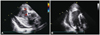
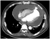
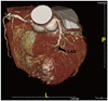
A 60-year-old man was a smoker and drinker with no history of coronary artery disease, hypertension and hyperlipidemia except diabetes, which was diagnosed several years ago. The man presented in the emergency room with complaints of chest pain for 2 days. The patient was conscious and physical examination showed heart rate 128 beats/min, blood pressure 95/70 mm Hg, temperature 36.5℃. Heart sound was faint without murmur and bruits. Initial electrocardiogram (ECG) showed atrial fibrillation and pathological Q waves in V 1-6. Chest X-ray revealed marked cardiomegaly. Blood tests were negative for Troponin I and creatine kinase-MB (CK-MB). Inflammatory markers were raised (white cell count of 23.12×10⁹/L, neutrophil percentage 88.4%, C reactive protein 16.84 mg/dL), and hemoglobin was mildly reduced (11.7 g/L). Kidney and liver enzymes were severely elevated (aspartate aminotransferase 2325 IU/L, alanine aminotransferase 1005 IU/L, γ-guanosine triphosphate 130 IU/L, lactate dehydrogenase 2989 IU/L, blood urea nitrogen 124.2 mg/dL, Creatinine 7.29 mg/dL). These lab results suggested that the patient was almost experiencing multiple organ dysfunction syndrome. Echocardiography showed reduced left ventricular (LV) systolic function and LV dilatation, especially at the LV apex. A large amount of pericardial effusion was noted. There was a small abnormal shunt from the LV apex to the pericardial space (Fig. 1). Thus, LVFWR was strongly suspected. In order to establish a diagnosis of LVFWR, chest computerized tomography (CT) was conducted and showed LV pseudoaneurysm with rupture in the anteroinferior wall with associated hemopericardium (Fig. 2). The patient underwent emergency LV reconstructive surgery (Dor procedure). The pericardial space was full of dirty fibrotic material. A small tear was found on the LV apex. The postoperative pathology revealed focal coagulation necrosis and abscess presented with blood clots and fibrin clots. Blood culture showed Streptococcus pneumonia. Postoperatively, intravenous antibiotic treatment was continued and the patient was discharged after 2 weeks with a good recovery. Three months later, coronary CT showed total occlusion of the mid left anterior descending coronary artery. Right coronary artery and left circumflex coronary artery was normal without stenosis (Fig. 3). At present, the aspirin therapy has been continued after discharge.
Discussion
Left ventricular free wall rupture is a dramatic complication of MI in which there is a rupture of infarcted LV free wall tissue. The rupture is commonly insidious with bleeding into the pericardial sac and subsequent cardiac tamponade.3) It is third to cardiogenic shock and arrhythmias as the leading cause of death following an MI.4) The most common location of LVFWR is in the anterior or lateral walls.5) It often appears within the first week following an acute MI, usually on the 4th or 5th day post-MI, although the time to manifestation may range from a few minutes to more than one month after the acute MI.6-9) Clinically, ruptures can be divided into 3 types: acute rupture, which results in death within a few minutes due to massive hemorrhage into the pericardial cavity; subacute rupture, which is characterized by a smaller tear that may temporarily be sealed by a clot or fibrinous pericardial adhesion and may be compatible with life for several hours or even longer; and chronic rupture with false aneurysm formation, which occurs when the leakage of blood is slow and when surrounding pressure on the epicardium temporarily controls the hemorrhage.10) The recent significant increase in the number of LVFWR diagnosed before death and the number of surgical repairs attempted is largely due to the widespread availability and use of cardiac imaging, particularly echocardiography.11) Echocardiography provides invaluable diagnostic information and indicates the possible extent and location of a rupture prior to surgery. The most frequent echocardiographic finding in the case of LVFWR is a localized pericardial effusion overlying the infarcted akinetic area. Other signs include echogenic 'specks' within the effusion and visible wall defects.12) In the current case, we found that pericardial effusion by echocardiography established the diagnosis of LVFWR by chest CT. On the basis of Troponin I, CK-MB and ECG, we thought that the anterior MI had occurred previously and led to LVFWR. At first, we had suspected coronary atherosclerotic heart disease was the reason of MI. In addition, we also could not rule out bacterial endocarditis, other sources of infection and embolization. The patient had diabetes (immunocompromised host) and atrial fibrillation. Atrial fibrillation could cause embolization (such as septic embolism) into coronary artery, then MI due to just embolism or abscess (caused by septic embolism) could occur without atherosclerotic coronary artery disease. The spread of infection affected the pericardial cavity and caused severe bacterial pericarditis. For this reason, CT angiography of the patient showed no evidence of coronary artery disease in other vessels except in the left anterior descending coronary artery occlusion. A pathologic report could not differentiate whether MI was atherosclerotic, or embolic, or secondary change according to abscess. The etiology of MI remains unclear.
In conclusion, rupture of the LV free wall is an often fatal complication of acute MI. Sometimes it presents insidiously. Physicians need to be aware of the risk factors, clinical signs, ECG and so on. This diagnosis can be made more accurately by using echocardiography and chest CT. Most of all, survival depends on early assessment of the patient, prompt investigation and diagnosis, and urgent surgical treatment.
 XML Download