

 PDF
PDF ePub
ePub Citation
 Print
Print


Introduction
Acute aortic dissection is an uncommon complication in patients with chronic hypertension. The separation of the layers within the aortic wall results in a false lumen with penetration of blood into the intima-media space. Many complications may ensue as the false channel enlarges, including myocardial infarction, acute heart failure, and rupture to neighboring structures.1) We report a patient who underwent successful surgical repair of an aortic dissection that had caused an aorta-right ventricular fistula.
Case
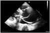
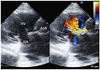
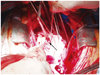
A 66-year-old woman presented to the emergency department of our center with complaints of melena and back pain in April 2010. The patient had a medical history that included hypertension which had been treated for 15 years with medication, an aortic valve replacement, and a MAZE procedure for degenerative aortic valve regurgitation 3 years prior. Her admission systemic blood pressure was 140/110 mm Hg, her pulse rate was 72 bpm, and her respiratory rate was 20 breaths per minute. A continuous murmur was noted at that time. A chest radiograph showed cardiomegaly with cardiothoracic index of 62%. Ventricular premature complex and left ventricular hypertrophy were noted on electrocardiogram. Esophagogastroscopy revealed no abnormal findings. Transthoracic echocardiography showed severely dilated aortic root and ascending aorta with intimal flap suggestive of aortic dissection (Fig. 1). Color Doppler demonstrated a shunt flow from aortic root to right ventricle, probably representing an aorto-right ventricular fistula (Fig. 2). The prosthetic aortic valve was normal. The left and right ventricles had normal function. Without further aortography or cardiac catheterization, we conducted emergency cardiac surgery. In the operation field, we found that the ascending aorta was severely enlarged. Although the prosthetic aortic valve was intact, the organized flap extended to the aortic root. A perivalvular adhesion was seen between the right atrium and the aortic wall, and a fistula had formed towards the right ventricle with less adhesion (Fig. 3). We conducted a modified Bentall operation with closure of the fistula. At 2 weeks post-surgery, echocardiography showed insignificant shunt flow from the ascending aorta to the right ventricle. The patient's condition improved and she was discharged 20 days after surgery.
Discussion
Acute aortic dissection is a life-threatening disease and may cause serious complications.2) Penetration into a cardiac chamber producing an aorto-cameral fistula is a very rare such complication. In 1924, Boyd3) reviewed 4000 autopsy reports of thoracic aortic aneurysm and found 1197 instances of rupture. The authors found several cases of fistula surrounding structures but no aortoright ventricular fistula. Although aortocameral fistulas have been rarely reported, the incidence tends to increase with previous cardiac surgery, particularly aortic valve replacement for aortic regurgitation.4) When aortic dissections occur in patients with previous cardiac surgery, postoperative adhesion may prevent free rupture into the pericardial space however these tend to penetrate into a neighboring cardiac chamber.5)6)
To diagnose a fistula into a neighboring structure found on aortic dissection, clinical history, physical examination, echocardiography, aortography, CT, and MRI may be helpful. Although aortography is the gold standard for diagnosis, non-invasive methods such as contrast enhanced CT, MRI, and echocardiography are currently preferred. Transesophageal echocardiography has better sensitivity and specificity than transthoracic echocardiography.7)8) Mortality in type A dissection after surgery is 26%, increasing up to 58% in cases that did not undergo surgery.9) Therefore, early diagnosis and immediate surgical intervention are important to reduce mortality in patients with aortic dissection. In this respect, transesophageal echocardiography is very helpful in aortic dissection enabling both diagnosis and detection of fistula by aortic dissection. CT, MRI, and aortography may be needed to confirm the diagnosis of aorto-cameral fistula, especially if echocardiography provides insufficient information. Although fistulization (aorto-right atrial and aorto-pulmonary) after acute dissection has been well documented, there are few reports of a fistula opening into the right ventricle.10-14)
Although aortic dissection complicated by a fistula is extremely rare, it should be suspected in any patient with a history of previous cardiac surgery, dyspnea, or murmur. In particular, echocardiography with Doppler is greatly helpful in checking the shunt caused by the fistula, as well as in visualization of the intimal flap.
 XML Download