

 PDF
PDF ePub
ePub Citation
 Print
Print


Introduction
Percutaneous coronary intervention (PCI) for chronic total occlusion (CTO) has a lower success rate than PCI for non-CTO lesions. A CTO lesion was defined as an obstruction of a coronary artery with thrombolysis in myocardial infarction or a flow grade of 0, with an estimated duration of at least 3 months prior to the event.
In 1990, the first retrograde approach technique was applied for CTO via a degenerated saphenous vein graft. Over the next 2 decades, septal collateral branches were considered to be novel access for the retrograde approach.
Variable and new retrograde techniques were reported over the next few years, including the following: kissing wire technique, knuckle wire technique, the controlled antegrade and retrograde subintimal tracking (CART) technique, the reverse CART technique, the modified reverse CART technique, the wire trapped technique, and the reverse wire trapped technique.
Despite the development of these various approaches, CTO lesion still remains a challenge. In this situation, information about the CTO lesion from intravascular ultrasound study (IVUS), cardiac CT angiography is a very useful approach, and is required for greater success in PCI.
In this case, we report our experience using the reverse CART technique under the aid of IVUS, cardiac CT angiography for an ambiguous CTO of proximal right coronary artery (p-RCA) (C, 100%, 0) with grade III collateral flow.
Case
A 68-year-old man with a history of hypertension presented with worsening exertional dyspnea. Fifteen months prior, angiography performed at a different hospital showed total occlusion of the p-RCA (100%, C, 0) and distal left circumflex artery (LCX) (100%, C, 0) with significant stenosis of the proximal left anterior descending artery (LAD) (95%, B2, II). At that time, PCI using drug-eluting stent was performed in the proximal LAD and distal LCX, only because multiple trials through the anterograde approach of CTO of p-RCA failed.
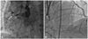
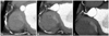
Bilateral coronary angiography demonstrated total occlusion of the p-RCA and rentrop grade 3 collateral flow from LAD, filling in a retrograde manner, the distal right coronary artery (RCA). There was no in-stent restenosis in proximal LAD and distal LCX. A small vessel of RCA proximal portion was looked as conus branch in coronary angiography (Fig. 1). But, IVUS study via anterograde approach in p-RCA revealed tapered-type CTO lesions, which was looked as conus branch. And, this distinction was detected in previous cardiac CT angiography, too (Fig. 2).
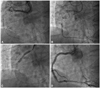
Therefore, we planned a retrograde RCA intervention. Multiple guide wires were used to bypass the lesion through use of the collaterals, retrospectively. Finally, the guide wire (Fielder FC 300 cm ASAHI INTECC, Osaka, Japan) passed the lesion via the septal branch to mid RCA. Then, a 2.5×15 mm size percutaneous transluminal coronary angioplasty balloon was inserted through the septal branch via retrograde guide wire and inflated to 12 atm. The second guide wire was introduced via anterograde approach. The lesion was dilated by 1.25×10 mm and a 2.5×15 mm balloon, respectively. Two cypher stents, 3.5×28 mm and 3.5×33 mm, were implanted in the mid- and proximal-RCA. Final angiography showed good distal flow without residual stenosis (Fig. 3).
Discussion
Percutaneous coronary intervention for CTO remains a challenge. Several studies show that successful recanalization of the CTO improved angina pectoris, survival, and left ventricular systolic function.1-5) Despite this necessity, the success rate of recanalization was still unsatisfied. The most common reason for PCI failure in CTO is passage failure of guidewire.6) IVUS can give adjunctive information to detect the area of occlusion in selective cases from adjacent side branches or from the false lumen and the anatomical information about the distal segment of the totally occluded coronary artery, when dye could not penetrate.7) In addition, cardiac CT angiography can help identify features that most influence the current success rates of PCI, such as marked calcifications at the stump, severe tortuosity of the proximal vessel, long length of the occluded segment, as well location of the vessel distal to the occlusion, which often cannot be well visualized using conventional angiography.8)
In this case, cardiac CT angiography and IVUS in addition to angiography were performed to evaluate CTO lesions because of the previous failure of intervention for p-RCA. Segmental calcified and soft plaques were detected in p-RCA with CTO by cardiac CT angiography. And, the conus branch was made out at the proximal portion of the plaques. Thus, we identified the lesion that firstly considered as tapered stump was conus branch. And it looked as tapered stump. Retrograde approach through the collateral channels has been proposed recently and has the potential to improve the success rate of PCI towards the treatment of CTO lesions.9) In addition, Rathore et al.10) demonstrated a 100% success rate of CTO recanalization using reverse CART.
In general, this technique is performed after previous techniques have failed via the anterograde approach, with the presumption that the distal cap of the CTO lesion may be softer than the proximal cap. Several strategies using the retrograde approach may be proposed to cross the occlusion, according to the CTO lesion characteristics, and sometimes, a very complex CTO lesion may require a combination of several techniques. The reverse CART technique has been shown to be safe and feasible, with a high success rate when performed by highly experienced operators.
In conclusion, if previous attempts to cross the totally occluded lesion from anterograde approach fail, the retrograde approach from collateral should be considered first, as in our case. With the help of diagnostic imaging, such as IVUS and cardiac CT angiography, it is possible to have more successful outcomes using PCI. In the future, the development of diagnostic methods will be reduced to reduce the failure to passage the CTO lesion.
 XML Download