

 PDF
PDF ePub
ePub Citation
 Print
Print


Introduction
Sarcoidosis is a multisystem granulomatous disease of unknown etiology. Cardiac manifestations, seen in approximately 5% of patients,1)2) typically include congestive heart failure with left ventricular dysfunction, conduction abnormalities, and ventricular arrhythmias. Moreover, sudden death has been reported in up to 67% of instances where cardiac sarcoidosis is diagnosed postmortem.3) Herein, we describe a patient whose cardiac sarcoidosis presented as symptomatic complete atrioventricular (AV) block with congestive heart failure and ventricular tachycardia. Tissue diagnosis was achieved via paratracheal node sampling, obtained by endobronchial ultrasonography guided transbronchial lymph node aspiration (EBUS-TBNA). The patient then received an implantable cardioverter-defibrillator (ICD) and systemic steroid therapy. Follow-up 18F-fluoro-2-deoxyglucose positron emission tomography (18F-FDG-PET) showed a marked decrease in cardiac sarcoid activity compared to the baseline.
Case
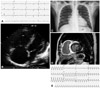
A 55-year-old male was admitted to another hospital six months previously for dyspnea on exertion (DOE) and dizziness. At that time, there was evidence of severe global hypokinesia of the left ventricle (LV), with systolic dysfunction {left ventricular ejection fraction (LVEF) 33% per M-mode} on echocardiogram, as well as enlargement of the right atrium and right ventricle (RV), and severe RV dysfunction. A corresponding electrocardiogram documented complete right bundle branch block with sinus bradycardia, while 24-hour Holter monitoring recorded a maximum RR interval of 2.76 seconds. No high-level AV block or arrhythmia was apparent, and by coronary angiography, epicardial coronary arteries were normal. On this basis, a diagnosis of idiopathic dilated cardiomyopathy was rendered and thereafter treated with ramipril 2.5 mg, furosemide 20 mg, and spironolactone 25 mg daily. Upon admission to our center, the patient again presented with DOE (NYHA class III) and intermittent dizziness, which were both aggravated in the preceding 15 days. His electrocardiogram showed complete AV block (heart rate, 27 beats per minute) (Fig. 1A) as did a 24-hour Holter monitor, with a maximum RR interval of 4.7 seconds. An echocardiogram demonstrated severe LV dysfunction (LVEF 34.4% per modified Simpson method), a dyskinetic basal septum, basilar cardiac akinesia, and an enlarged, severely dysfunctional RV, without pulmonary hypertension {pulmonary artery systolic pressure (RVSP) 27.64 mm Hg} (Fig. 1B). Mild cardiomegaly was noted on chest X-ray (cardiothoracic ratio 55%), but hilar lymph nodes were not prominent (Fig. 1C). Gadolinium-dietylene triamine penta-acetic acid (DTPA)-enhanced cardiac MRI showed delayed hyper-enhancement of the entire RV wall and subepicardial (apical) or transmural (apical, basal) portions of the LV (Fig. 1D).
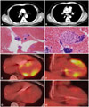
While cardiac sarcoidosis was strongly suspected, endomyocardial biopsy findings were nonspecific, and angiotensin-converting enzyme (ACE) was within normal reference range (27.5 IU/L; normal range: 8.0-55.0 IU/L). During endomyocaridal biopsy, sustained ventricular tachycardia developed repetitively (Fig. 1E). Enlarged right paratracheal (17.27×8.35 mm) and subcarinal (18.63×11.97 mm) lymph nodes, found incidentally with coronary CT angiography six months prior (Fig. 2A and B, respectively), were instead sampled via EBUS-TBNA to histologically confirm the presence of noncaseating epitheloid granulomata with giant cells (Fig. 2C and D). In accordance with the Japanese Ministry of Health and Welfare guideline,4) cardiac sarcoidosis was diagnosed and a systemic glucocorticoid (prednisolone, 60 mg daily) was initiated. An ICD was also inserted as a precautionary measure for ventricular tachycardia and symptomatic complete AV block. Because the ACE level was within the normal reference range, we used 18F-FDG-PET to monitor therapy. Baseline results at >18 hours prolonged fasting showed inhomogeneous 18F-FDG uptake, accentuated in LV myocardium {maximum standardized uptake value (SUVMAX)=7.9 g/mL} (Fig. 2E and F). On discharge, the patient was asymptomatic. One month later, follow-up 18F-FDG-PET showed a marked decrease in myocardial hypermetabolism (SUVMAX=3.1 g/mL) (Fig. 2G and H), and the right paratracheal lymph node had decreased in size upon chest CT. The ACE level also dropped to 14.1 U/L. The patient is now doing well, without recurrent symptoms, including ventricular tachycardia.
Discussion
Sarcoidosis is a multisystem disease that has a worldwide distribution, characterized by the presence of noncaseating granulomata in involved organs. Its etiology is currently unknown. The prevalence of this disease varies from 10 per 100000 in white populations to 35 per 100000 in African Americans.5-7) In the US, around 20-27% of patients with sarcoidosis have myocardial involvement,8) while in Japan, up to 58% of patients have cardiac lesions, accounting for as many as 85% of recorded deaths from sarcoidosis.9)
Reports also indicate that cardiac involvement is largely unrecognized antemortem. In only about 5% of patients with autopsy evidence of cardiac sarcoid (i.e., noncaseating granulomata) is cardiac disease apparent prior to death.10) Common presentations of cardiac sarcoidosis include congestive heart failure with left ventricular dysfunction, cardiac rhythm disturbance, and most importantly sudden death. The latter has been cited in up to 67% of patients where cardiac sarcoidosis was diagnosed postmortem.3)
Among the available noninvasive diagnostic modalities, gadolinium-DTPA-enhanced cardiac MRI offers greater utility for early diagnosis and follow-up, compared with the low-sensitivity of endomyocardial biopsy (approaching 20%).11-13) Recent studies have similarly demonstrated the potential of 18F-FDG-PET as a diagnostic and follow-up tool.14-16) Because the ultimate goal for these patients is to preserve cardiac function and prevent fatal arrhythmias, early detection, even in absence of symptoms, is critical. In this respect, gadolinium-DTPA-enhanced cardiac MRI and 18F-FDG-PET may more reliably facilitate early diagnosis and thus result in better treatment outcomes.14) In our patient, delayed myocardial enhancement by cardiac MRI was a key diagnostic clue. Despite procurement of an adequate endomyocardial biopsy, a tissue diagnosis could not be established with this approach. It was through EBUS-TBNA of right paratracheal and subcarinal lymph nodes that the diagnosis of cardiac sarcoidosis was eventually made.
Systemic steroids have shown survival benefits with cardiac sarcoidosis and have been generally accepted as the treatment of choice17)18) However, the effect of steroids on ventricular arrhythmias is less certain, and some recommend an ICD as primary therapy in patients presenting with ventricular tachycardia.10)19) In addition to a systemic steroid, our patient received an ICD to deal with symptomatic complete AV block and ventricular tachycardia.
Serum ACE level typically reflects sarcoid activity and can be used as a biomarker, although our patient's levels were normal. Furthermore, cardiac MRI was precluded for therapeutic monitoring, due to the precautionary ICD, so 18F-FDG-PET was used. At the one-month interval, myocardial hypermetabolism had markedly decreased by 18F-FDG-PET, which correlated well with the patient's clinical response and a corresponding decline in his serum ACE level.
In conclusion, cardiac sarcoidosis presenting with symptomatic complete AV block and sustained ventricular tachycardia, was successfully treated in this instance with a systemic steroid and an ICD, while serum levels of ACE and 18F-FDG-PET aided in therapeutic monitoring.
 XML Download