

 PDF
PDF ePub
ePub Citation
 Print
Print


Introduction
Pericardial abscess is a rare condition which results from hematogenous spread, direct extension from an adjacent infectious focus, trauma, or surgery. A pericardial abscess by Staphylococcus aureus is rarer1-3) and to our knowledge, this is the first case report of a pericardial abscess as a complication of staphylococcal bacteremia in Korea.
Case
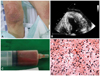
A 72-year-old woman presented to the emergency room with dyspnea and myalgia that developed 2 weeks after undergoing acupuncture therapy on both knees due to arthralgia. Vital signs on admission showed hypotension (80/40 mm Hg), tachycardia (118 beats per minute), tachypnea (20 per minute), and hypothermia (35℃). On physical examination, the patient had multiple needle scars on both knees with dappled rashes on her entire body (Fig. 1A). Her laboratory results showed elevated white blood cell counts (10100/mm3), with neutrophil 88%, elevated high sensitive C-reactive protein level of 36 mg/dL (reference range, 0-0.5 mg/dL), blood urea nitrogen/creatinine 71/4.5 mg/dL, myoglobin 5169 ng/mL (reference range, 16.3-96.5 ng/dL), creatine kinase myocardial band 9.7 U/L (reference range, 0-3.6 U/L), Troponin I 0.28 ng/mL (re-ference range, 0-0.1 ng/mL). A chest radiograph showed cardiomegaly and an electrocardiography showed atrial fibrillation with rapid ventricular response. A transthoracic echocardiogram demonstrated concentric left ventricular hypertrophy with fluid collection in the posterolateral wall of the pericardium with no evidence of valvular vegetation or tamponade physiology (Fig. 1B). Pericardial aspiration of the fluid revealed a bloody material (Fig. 1C) and cultures grew Staphylococcus aureus. Blood cultures showed staphylococcal bacteremia (Fig. 1D). Lab analysis of aspiration fluid showed elevated white blood cell counts >50000/mm3), with polymorph-onuclear neutrophil 90%, pH 7.3, glucose 5 mg/dL, lactate dehydrogenase 12397 U/L, albumin 2.4 g/dL and total protein 6.2 g/dL. Percutaneous drainage and empiric antibiotic treatment were started immediately. However, the patient expired due to refractory sepsis and organ failure.
Discussion
Pericardial abscess is a serious, life-threatening illness associated with high mortality. A pericardial abscess is an extremely unusual complication of Staphylococcus aureus bacteremia.1-4)
The mechanism of purulent pericarditis by Staphylococcus aureus is unknown. Possible explanations include hematogenous seeding or direct extension into a pre-existing pericardial cyst or purulent pericarditis occurring in a patient with old pericardial adhesions.5)6) Other microorganisms causing pericardial abscess include Mycobacterium tuberculosis, Gram-negative bacilli, Streptococcus species, and Aspergillus. In Korea, only two cases of tuberculous pericardial abscess and Bacteroides fragilis have been reported.7)8)
Because delayed diagnosis of pericardial abscess may lead to debilitating complications, early echocardiography is important. To-mography provides useful information on the extent of the pericardial abnormality when the echocardiographic picture is not clear.5) The primary treatments for pericardial abscess include percutaneous or surgical drainage and pericardiectomy with prompt administration of appropriate antibiotics.
Although we cannot verify the pathogenesis of this patient's infection, based on the multifocal acupuncture therapy history of this patient and the absence of previous pericardial disease, the pericardial abscess may have been caused by hematogenous spread of Staphylococcus aureus from the soft tissue infection of the knees. However, it remains to be determined whether acupuncture treatment severely increases the risk of bacteremia, or whether this case is simply a coincidence implicating acupuncture.
 XML Download