

 PDF
PDF ePub
ePub Citation
 Print
Print


Introduction
The presence of myocardial ischemia causes various symptoms in patients and is predictive of future events1)2) and revascularization of those lesions is important since it has the potential to improve patient outcomes.2-4) However, revascularization of stenotic lesions that do not lead to myocardial ischemia is not beneficial and can rather be harmful. Therefore, the decision to revascularize a coronary artery stenosis should be guided by the evidence of myocardial ischemia.
Coronary angiography is limited in its ability to determine the physiologic significance of coronary stenosis.5)6) Especially in patients with intermediate stenosis, angiographic information does not correlate well with the functional significance of a lesion.7-9) This uncertainty may result in unnecessary revascularization of insignificant lesions or failure to revascularize the clinically significant ones. As a result, fractional flow reserve (FFR) was introduced and has proven to be a reliable method for determining the functional significance of coronary stenosis. FFR expresses the maximal achievable blood flow in a coronary vessel as a fraction of normal maximal blood flow to the same myocardial territory.10) In other words, FFR represents the extent to which maximal myocardial blood flow is limited by the presence of epicardial stenosis and can be easily measured by the ratio of distal coronary pressure to aortic pressure during maximum hyperemia (Fig. 1). This index is independent of changes in hemodynamic conditions such as systemic blood pressure, heart rate, or myocardial contractility.11) As the clinical benefit of an FFR-guided revascularization strategy has been proven in several studies with different lesion subsets, this strategy has become more popular in recent years (Fig. 2).
Fractional Flow Reserve: The Past
In the very early period of percutaneous coronary intervention (PCI), clinical application of intracoronary pressure was tried in patients with coronary artery stenosis but failed. However, the cause of failure at that time was due to the fact that intracoronary pressure was measured with a large over-the-wire balloon catheter without hyperemia (minimal microvascular resistance). Since then, clinical application of intracoronary pressure had been almost forgotten until the concept of myocardial FFR was developed and introduced by N. Pijls and B. De Bruyne in the early 1990s.
The concept was first validated in an animal study12) and later in humans using a positron emission tomography scan.13) Given that FFR is a continuous variable, a certain cutoff value was necessary to determine the presence of myocardial ischemia (dichotomous variable). In 1996, Pijls et al.10) performed a clinical study to define the cutoff value of FFR to determine the presence of ischemia using non-invasive tests and sequential Bayesian considerations. In this study, an FFR cutoff value of 0.75 had a positive predictive value of 100% and a negative predictive value of 88% to determine the presence of ischemia. Due to a small zone of uncertainty between 0.75 and 0.80 (grey zone) and the results of the FFR versus Angiography for Multivessel Evaluation (FAME) study,3) many clinicians now use the FFR cutoff value of 0.80 as a guide to perform revascularization.
After validation of a cutoff value, the clinical benefit of FFR-guided revascularization was tested in the DEFER study (FFR to Determine the Appropriateness of Angioplasty in Moderate Coronary Stenoses).4) This study included 325 patients referred for PCI of a single, de novo stenosis of intermediate severity. PCI was performed in all patients with an FFR <0.75 (reference group, n=144). If the FFR was ≥0.75, patients were randomized to either medical treatment (defer group, n=91) or PCI (perform group, n=90). After 5 years of follow-up, event free survival did not differ between the defer and PCI groups (80% and 73%, respectively) and the percentage of patients free from chest pain at follow-up was not different between the 2 groups. The composite rate of death and acute myocardial infarction (MI) in the defer group was only 3.3% during the period of 5 years. This study showed that patient outcomes with deferral of PCI according to FFR was excellent and the risk of death or acute MI was <1% per year which could not be further decreased by stenting. Since then, the benefit of FFR-guided revascularization strategy was tested and confirmed in more complex scenarios involving multiple lesions, multivessel disease, in-stent restenosis, post-stenting, left main disease, bifurcation lesions and patients with MI.14-19) These results culminated in a Class IIA recommendation of FFR in 2007 American College of Cardiology/American Heart Association Society for Cardiac Angiography and Interventions PCI Guidelines on myocardial revascularization: "It is reasonable to use intracoronary physiologic measurements (Doppler ultrasound, fractional flow reserve) in the assessment of the effects of intermediate coronary stenoses in patients with angina symptoms" (Table 1).20)
Fractional Flow Reserve: The Present
A nuclear substudy of the Clinical Outcomes Utilizing Revascularization and Aggressive Drug Evaluation trial showed that PCI could improve the outcome of patients with coronary artery disease (CAD) which resulted in the relief of myocardial ischemia.2) Investigators of the FAME study addressed the hypothesis that an FFR-guided PCI approach with drug-eluting stents would be superior to the current practice of conventional angiography-guided PCI in patients with multivessel CAD. The FAME protocol directed the investigators to stent a lesion with at least 50% stenosis and if the investigators thought that stenting was warranted on the basis of available clinical information. The patients were then randomized 1 : 1 to either standard PCI as planned (n=496) or to FFR-guided PCI (n=509). Although the number of angiographically significant stenoses was identical between the 2 groups (2.7±0.9 vs. 2.8±1.0), the FFR group used fewer stents per patient (1.9±1.3 vs. 2.7±1.2, p<0.001) and less contrast medium (272 mL vs. 302 mL, p<0.001). More importantly, at 1-year follow-up, the FFR group had fewer total cli-nical events (13.2% vs. 18.4%, p=0.02) and fewer combined death or MI (7.3% vs. 11%, p=0.04) compared to the angiography-guided PCI group. At 2 years, the rate of combined mortality or MI was still in favor of the FFR group (8.4% vs. 12.9%, p=0.02).21) Further analysis showed that an FFR-guided strategy is not only cost-effective but also cost-saving compared to an angiography-guided strategy.22) Another important finding of the FAME study is that assessment by FFR in patients with multivessel disease can lead to a reduction in the number of diseased coronary arteries and change in the treatment strategy. Of all patients with angiographic triple vessel disease (VD) in the FFR group, only 14% of the patients had functionally significant triple VD and 86% had ≤2 functionally significant diseased coronary arteries (2-VD=43%, 1-VD=34%, 0-VD=9%).8) Furthermore, the functional SYNTAX score (SYNTAX score only by ischemia-inducing lesions as determined by FFR) was shown to decrease the number of high-risk patients and better discriminate the risk for future adverse events in patients with multivessel CAD.23)
Fractional Flow Reserve: The Future
Although FFR has become the gold standard invasive assessment to detect the ischemia-related lesion, it requires an invasive procedure, expensive devices and pharmacologic intervention to induce maximal hyperemia. Therefore, further development is still necessary to expand the clinical applications of FFR.
Novel hyperemic stimuli
Continuous infusion of adenosine via the central vein has been considered as the gold-standard method of hyperemia for FFR measurement.25) However, this method requires relatively large doses of adenosine resulting in high cost, an additional procedure for femoral vein access and is practically not feasible during transradial coronary catheterization procedures. Furthermore, the adenosine administration itself is associated with adverse systemic effects such as AV block, dyspnea and chest pain.26)27)
To overcome the complexity of central vein infusion of adenosine, the feasibility and efficacy of peripheral infusion of adenosine were tested in recent studies.28)29) Seo et al.29) compared the hyperemic efficacy between continuous IV infusion (140 µg/min/kg) via the femoral vein and the forearm vein and found that the hyperemic efficacy of the forearm vein infusion (FFR: 0.80±0.11) was not inferior (p for non-inferiority=0.01) to the femoral vein infusion (FFR: 0.80±0.10) of adenosine. The number of functionally significant stenoses was not different between the 2 methods {femoral vein vs. forearm vein; 17 (25.0%) vs. 17 (25.0%), p=1.0}. Therefore, this method can be used for FFR measurement, especially during transradial coronary catheterization procedures.
Novel hyperemic agents for invasive physiologic assessment were also introduced. Nair et al.30) compared the hyperemic efficacy between a selective A2A receptor antagonist, regadenoson (400 ug, IV bolus) and adenosine in 25 patients with intermediate coronary stenosis and found that a single IV bolus of regadenoson was as effective as an IV infusion of adenosine. Jang et al.31) compared the hyperemic efficacy of a bolus administration of nicorandil (intracoronary, 2 mg) with continuous infusion of adenosine in 210 patients. In this study, hyperemic efficacy of nicorandil was not inferior to that of adenosine (0.82±0.10 vs. 0.82±0.10; p for non-inferiority<0.001) and there was a strong linear correlation between the FFR measured by IV infusion of adenosine and nicorandil (R2=0.934). Moreover, nicorandil caused less changes in mean blood pressure, heart rate, PR interval and less severe chest pain than adenosine (p<0.05). While transient AV block occurred in 16 patients with adenosine, none were detected with nicorandil.
These novel agents and methods of adenosine administration will cause less discomfort in patients and reduce the complexity of invasive physiologic assessment.
Novel physiologic index without hyperemia
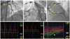
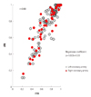
A new physiologic index, instantaneous wave-free ratio (iFR) without the requirement for hyperemia was introduced and tested in Adenosine Vasodilator Independent Stenosis Evaluation (ADVISE) study.32) From the meticulous investigation on coronary flow and resistance, the investigators found that there is a certain period in the cardiac cycle during which the resistance at rest is similar in variability and magnitude to those during hyperemia. In the ADVISE study, the distal-to-proximal pressure ratio during this period, which is also known as iFR, was compared with FFR in 157 stenoses. In this study, iFR had a good correlation with FFR (r=0.9, p<0.001) with excellent diagnostic performance (Fig. 3). This novel concept, iFR, has great appeal as it may provide a faster and easier invasive physiologic assessment for CAD. However, this concept still awaits further validation.
Non-invasive assessment of fractional flow reserve using CT scan
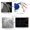
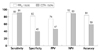
Recent advancements of CT technologies have enabled several novel methods to assess the functional significance of coronary stenosis in addition to anatomical information. One of these is the application of computational fluid dynamics to coronary CT angiography (CCTA) images.33) With this technology, FFR can be computed using images from conventional CCTA (CT-derived computed FFR; FFRCT) without any invasive procedure and without hyperemia (Fig. 4). A prospective, multicenter clinical trial, Diagnosis of Ischemia-Causing Stenoses Obtained Via Noninvasive Fractional Flow Reserve (DISCOVER-FLOW) study, was performed to assess the diagnostic performance of FFRCT in the prediction of the functional significance of stenosis.34) In this study, 103 patients (159 vessels) with stenosis in a major epicardial coronary artery who had diagnostic quality CT images from 64 or more detector row CT scanners were consecutively enrolled and the diagnostic accuracy of CCTA (≥50% stenosis) and FFRCT were compared. On a per-vessel basis, accuracy, sensitivity, specificity, positive predictive value, and negative predictive value for FFRCT and CCTA were 84.3%, 87.9%, 82.2%, 73.9%, 92.2%, respectively, and 58.5%, 91.4%, 39.6%, 46.5%, 88.9%, respectively (Fig. 5). This study showed that noninvasive FFR derived from CCTA (FFRCT) had a high diagnostic performance for the detection and exclusion of coronary lesions that lead to ischemia. Clinical application of this novel technology may potentially reduce unnecessary invasive procedures. Moreover, combination of virtual intervention and this technology can help to determine the treatment strategy in complex lesions prior to the invasive procedure. Although the concept and results of the DISCOVER-FLOW study are very encouraging, further studies with a larger number of patients are needed to validate the clinical usefulness of this novel technology. A larger, prospective multicenter clinical trial, Determination of Fractional Flow Reserve by Anatomic Computed Tomographic Angiography study, has completed patient enrollment and the results will soon be available.35)
Conclusion
Fractional flow reserve has become the gold standard to define the functional significance of coronary stenosis. Novel hyperemic stimuli and novel physiologic indices without hyperemia will reduce the barriers and expand the clinical application of FFR. Furthermore, non-invasive assessment of the functional significance of coronary stenosis such as CT-derived computed FFR, can be helpful in optimizing the interventional treatment strategy for patients with CAD prior to the invasive procedure.
 : hyperemic myocardial blood flow in the presence of a stenosis,
  : normal hyperemic myocardial blood flow, Pd: distal coronary pressure, Pa: aortic pressure, Pv: venous pressure, R: hyperemic myocardial resistance.
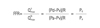

 XML Download