

 PDF
PDF ePub
ePub Citation
 Print
Print


Introduction
Single anomalous origin of coronary artery is a rare congenital anomaly of coronary circulation.1) The incidence of single anomalous origin is approximately 1% among patients undergoing cardiac catheterization, and an anomalous origin of the left coronary artery is particularly less frequent than the right coronary artery (RCA).2-7) We report on a rare case of anomalous left coronary artery that originated from the right sinus of valsalva, with total occlusion on the proximal RCA.
Case
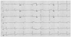
A 49-year-old male suffered from chest pain, dyspnea as well as hypotension and pulmonary congestion 10 hours prior to arrival at the emergency room. Risk factors included smoking and dyslipidemia. The electrocardiogram (ECG) showed ST-segment elevation in aVR and V 1-2, and ST-segment depression in II, III, aVF and V 4-6 (Fig. 1), and troponin-I was elevated (1.35 ng/mL). Under the diagnosis of non-ST segment elevation myocardial infarction, the patient was immediately sent to the catheterization laboratory.
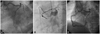
The first time, a coronary angiogram (CAG) suspected an anomalous origin of left coronary arteries and suspected right ventricular branch communicating between proximal RCA and middle left anterior descending coronary artery (LAD) (Fig. 2A), and revealed thrombotic total occlusion in proximal RCA (Fig. 2B). Therefore, for RCA lesion, multiple stepwise plain old balloon angioplasties (POBA) with 1.5×13 mm and 2.5×15 mm balloons, and multiple thrombi suctions with Thrombuster® (Kaneka Corporation, Osaka, Japan) were performed. During the procedure, a temporary pacemaker was inserted due to junctional bradycardia and deepening of mental status. Although multiple thrombi were extracted, huge thrombi remained in distal RCA. Therefore, additional POBA and thrombosuction were performed repeatedly, and intracoronary glycoprotein IIb/IIIa receptor blocker (abciximab) was injected in RCA, after which a 4.0×25 mm bare-metal stent (Coroflex blue®, Braun, Berlin, Germany) was deployed in critical fixed stenosis of the proximal RCA. The RCA showed good distal flow and markedly decreased residual stenosis (Fig. 2C).
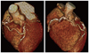
The echocardiography noted mild left ventricular systolic dysfunction (EF=43.9% by biplane method), and akinesia in RCA and left circumflex artery (LCX) territory. Cardiac computed tomography (CT) scan showed high calcium score (2036.49) and single origin of coronary artery from right coronary sinus of valsalva with communication between RCA and middle LAD via right ventricular branch (Fig. 3), and moderate degree of luminal narrowing with mixed plaque in middle LAD, and hypoplastic LCX. We maintained IV heparin for the resolution of thrombi on admission.
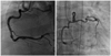
One week later, we performed follow-up CAG using Amplatz right 2 cm curve (AR2) guiding catheter, CAG revealed patent stent in proximal RCA and a more resolved state of thrombi in PDA, as well as improved distal flow in the posterolateral branch (Fig. 4). After uneventful recovery, the patient was discharged with dual antiplatelet therapy (aspirin 100 mg, clopidogrel 75 mg) and has been followed up at the outpatient clinic.
Discussion
Anomalous origin of coronary arteries is a rare congenital anomaly that was first described in 1948 by White and Edward.1) The prevalence of this anomaly was determined as 0.6% to 1.3% in an angiographic series and 0.3% in autopsy series.2-6) In particular, incidence of a left coronary anomaly that originates from RCA has been reported to be very rare (0.016% incidence).7) In Korea, a few cases of left coronary anomaly were reported, and most of them were left coronary artery that originated from the right coronary sinus.8,10) Unlike the previous cases, the present case showed a single origin of coronary artery from the right coronary sinus of valsalva with communication between RCA and LAD coronary artery via the right ventricular branch.
Manifestations vary according to subtype of anomalous origin from asymptomatic patients to those who present with myocardial ischmia, angina pectoris, arrhythmia, syncope, and also sudden death, in absence of atherosclerosis.11)12) The pathophysiology basis is unclear. The restricted coronary blood flow in this anomaly suggests that the acute takeoff angle, slit-like orifice, and compression of the intramural segment by the aortic valve commissure are considered to narrow the orifice.13)
The correlation between coronary artery disease and coronary anomalies is uncertain.13) Some previous literature data suggested that anomalous origin of the coronary artery could make them more inclined to atherosclerosis because of altered blood flow pattern.14)15) However, the incidence of coronary artery disease was no different compared to normally-originating coronary arteries.16)17)
The screening method used to evaluate anomalous coronary arteries is important, because this anomaly can be associated with ischemic heart disease and sudden cardiac death. Instrumental advances in multi-detector CT (MDCT) have realized the reduction of imaging time, dose of contrast medium, and radiation exposure. MDCT can be used for non-invasive diagnosis of anomalous origin of coronary arteries, and be useful in increasing the success rate of cannulation during CAG.
Since manifestation varies from benign to sudden death depending on situation, multiple treatment options should be considered including medical, interventional, or surgical procedures.18) Recently, a report suggested that an anomalous coronary artery should be followed without intervention and stated that the benefit of excessive exercise limitation is doubtful. If a young, symptomatic patient has significant luminal narrowing on imaging studies, surgical intervention should be considered.13)19)
In this case, we report a patient with anomalous left coronary artery that originated from the right sinus of valsalva accompanied with total occlusion on the proximal RCA, who underwent successful percutaneous coronary intervention.
 XML Download