

 PDF
PDF ePub
ePub Citation
 Print
Print


Introduction
Coronary artery anomalies (CAA) are rare angiographic findings. The incidence of CAA is about 1-2% in angiographic studies of the adult population.1) Double right coronary artery (RCA) is a very rare coronary anomaly. Patients are mostly asymptomatic but acute coronary syndrome (ACS) is a possible, yet uncommon clinical presentation. ACS is linked to CAA in some cases.
We report a rare anomaly, an A2 atypical double RCA, which presented as an acute inferior wall myocardial infarction due to a thrombotic occlusion in one of the two RCAs, which was successfully managed with primary percutaneous coronary intervention (PCI).
Case
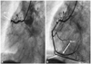
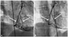
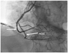
A 61-year-old male patient was admitted to the emergency department with retrosternal chest pain at rest for the past hour. He was a chronic smoker and had hypertension for 5 years. Blood pressure was 130/80 mm Hg, and pulse rate was 86 beats/minute. A physical examination was completely normal. An ST segment elevation was noted in leads II, III, and aVF; and a reciprocal ST segment depression in leads V 1-6 on electrocardiography. Coronary angiography showed total occlusion of the proximal RCA (Fig. 1A). The origin and course of the left coronary arteries were normal. Noncritical lesions were evident in the left anterior descending artery and circumflex artery. Primary PCI was performed on the RCA lesion. The lesion was predilated with a 2.0×15 mm balloon at 12 atm. Two different RCAs originating from a common main trunk appeared following a contrast injection to the right coronary orifice (Fig. 1B). Tubular eccentric stenosis of 90% was noted in the proximal segment of the main RCA trunk, 70% tubular stenosis in the RCA1, and 40% stenosis in the osteal segment of RCA2 (Fig. 2A). Two bare metal stents (3.0×18 mm and 2.75×12 mm) were successfully deployed to the lesion at 15 atm for 30 seconds in the proximal segment of the main trunks of RCA and RCA1, respectively (Fig. 2B). A control coronary angiogram demonstrated two separate RCAs originating from a single ostium. So, we diagnosed the patient with an A2 atypical double RCA (Fig. 3). The patient was treated with a tirofiban infusion. The post-interventional period was uneventful, and the patient was followed up for 2 days at the intensive care unit and discharged without any complications.
Discussion
Most coronary anomalies are detected as incidental findings during coronary angiography. However, a double RCA is a very rare coronary anomaly. So far, only a few cases of double RCA have been reported.2-5) Since the second report, this rare anomaly has been accepted as a benign entity. However, new reports in the last few years have indicated that a double RCA may be complicated with atherosclerosis. Among 27 cases defined in the literature, atherosclerosis was identified in eight patients; seven presented with ACS, and three with inferior myocardial infarction.6)7) In most of the reported double RCA cases with atherosclerosis, the atherosclerotic segment is usually in the proximal part of the RCA that courses on the atrioventricular (AV) sulcus or in the posterior branch of the RCA going to the AV sulcus.8) In the submitted case of Akçay et al.9) and in our case, a lesion was evident in the ostial part of the anteriorly coursing RCA but not in other parts. Also, in their case, an atherosclerotic lesion was found in the posterior course of the RCA. Based on these two studies by Iacobellis et al.10) the authors suggested that an increased quantity of epicardial adipose tissue due to obesity may be a risk predictor for cardiovascular disease.
An attempt to classify a double RCA was tried (Table 1) by an author from Turkey, where most of the studies concerning this anomaly have been reported.11) We diagnosed our case as an A2 atypical double RCA using this classification. Only one case has been reported that was admitted with ACS and identified as atypical RCA by coronary angiography.11) In that case, atherosclerotic lesions were only found in the proximal part of the RCA but not in the RCA1 or RCA2, and the stent was implanted in the main RCA. But in our case, atherosclerosis began proximally and was ongoing in RCA1 and RCA2, so we implanted stents proximal to RCA and RCA1.
To our knowledge no A2 atypical double RCA case has been reported that was admitted with ACS and had atherosclerosis in the main RCA as well as in RCA1 and RCA2.
In conclusion, although controversy exists about the definition of a double RCA, it is generally considered a benign entity, but could be atherosclerotic and cause ACSs including myocardial infarction and may be associated with other anomalies.12)13) Coronary anomalies should be recognized to avoid problems during coronary intervention and cardiac surgery. Every operator should be familiar with coronary anomalies to perform an adequate examination.

 XML Download