

 PDF
PDF ePub
ePub Citation
 Print
Print


Introduction
The safety of contrast agent in echocardiography was reported and there is a study showing that these agents have a good safety profile in both cardiac and abdominal ultrasound applications.1) Here we introduced a simple diagnostic approach to coronary artery fistula with contrast agent during echocardiography and it helped us to access the diagnosis because of the typical diastolic flow in pulmonary artery.
Case
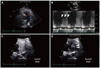
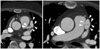
A 46-year-old female was admitted to the hospital with progressive dyspnea. She had been suffering from dyspnea on exertion for 5 years but could not be diagnosed. No abnormal findings were observed on an initial chest posterioaneterior view and electrocardiogram. However cardiac echocardiography revealed abnormal small turbulent flow in the main pulmonary artery (Fig. 1A), and Doppler revealed diastolic-dominant flow (Fig. 1B). Because there was no evidence of a right-to-left shunt, pulmonary parenchymal disease, or hypersensitivity to perflutren, which are contrast agent contraindications, we administered the Definity® contrast agent (Bristol-Myers Squibb Medical Imaging, North Billerica, MA, USA) in real echocardiographic mode (infusion rate of 3.0 mL/min mixed with normal saline, 50 mL) using a Vivid 7 (GE Ultrasound, Horten, Norway) ultrasound system and found unusual whitish flow in the main pulmonary artery during the diastolic phase (Fig. 1C and D). Under the impression that this was a coronary artery fistula, we performed aortic computed tomography (CT) and revealed two huge right coronary artery fistulas in the main pulmonary artery (Fig. 2). Finally she underwent a surgical correction.
Discussion
Coronary artery fistulas account for only 0.4% of congenital heart defects2) and approximately 50% of pediatric coronary vasculature anomalies. The incidence of coronary artery fistula in the overall population is estimated to be about 0.002%, and 20% of patients with a congenital coronary artery fistula have other concomitant car-diac anomalies, most frequently aortic and pulmonary atresia and patent ductus arteriosus. Tetralogy of Fallot has also been reported.3-5) Congenital fistulas often arise from the right coronary artery system and the majority enter the right ventricle, right atrium, superior vena cava, coronary sinus, or pulmonary arteries.6)
Many patients are asymptomatic; however, an awareness of these fistulas is important because they have been associated with various clinical features including chest pain or heart failure in young patients.7) Coronary artery fistulas have been diagnosed with aortography,8) coronary angiography,9) and coronary CT.7) Although there is a case of colour Doppler assessment of a coronary fistula,10) we cannot easily confirm these fistulas with echocardiography. In this study, we used a simple diagnostic approach for a coronary artery fistula using a contrast agent, which aided with the diagnosis, because of the typical diastolic whitish flow in the pulmonary artery. The safety of the contrast agent (including Definity®) has been reported, and Wei et al.1) showed that these agents have a good safety profile in both cardiac and abdominal ultrasound applications. The incidence of severe adverse reactions to ultrasound contrast agents is no greater and may be lower than that reported for contrast agents commonly used in other cardiac imaging tests.1) The flow of a coronary artery fistula can be noted easily during the diastolic phase by contrast infusion, so the coronary artery fistula can be approached in a more direct way.
 XML Download