

 PDF
PDF ePub
ePub Citation
 Print
Print


Introduction
Totally implantable central venous access devices for administration of chemotherapeutic agents (chemoport) are commonly used in cancer patients with an indication for chemotherapy. Percutaneous implantation of a chemoport via the subclavian vein is associated with several kinds of potential complications, including pneumothorax, malposition, infection, bleeding, arrhythmias and thrombosis, both during placement and later in long-term maintenance.1) Transection and embolization of a chemoport, so called "Pinch-off" syndrome (POS), is a rare complication and the reported incidence is approximately 1.6 percent (26/1644), with a range of 0.1 to 2.1 percent.2)
We present a case in which a chemoport embolized into the inferior vena cava (IVC) and was successfully retrieved by a percutaneous approach using a goose neck snare.
Case
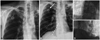
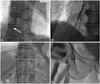
A 66-year-old man presented with descending colon cancer (T3N1 M0, stage IIIA) in 2007. The lower anterior resection of colon and adjuvant chemotherapy was performed for this patient, and he was doing well. He performed regular imaging and medical check up after the treatment. Non-symptomatic metastatic nodule was discovered in follow up abdominal computed tomography on 2 years after the surgery. He underwent segmentectomy of the liver and an implantable central venous access device, the chemoport (Celsite ST 201, 8.5 Fr, B. Braun Medical Inc, Melsungen, Hesse, Germany), was placed in the right subclavian area, for administration of chemotherapeutic agent. The post-insertion chest X-ray (CXR) confirmed good placement of the chemoport (Fig. 1A). Six-days after implantation of the chemoport, he complained of chest discomfort and swelling of right infraclavicular area. A CXR showed that the distal portion of the chemoport had been fractured and embolized into the IVC (Hinke Grade 3) (Fig. 1B). He was transferred to the cardiac catheterization room for percutaneous retrieval of embolized part of chemoport. Puncture of the femoral vein was followed by insertion of a 7 Fr sized venous sheath. The fractured distal segment of the chemoport was subsequently caught and moved into the femoral vein using a goose neck snare (vascular retrieval forceps/3 Fr/120 cm, Cook Inc, Bloomington, IN, USA) (Fig. 2). The distal segment of the chemoport was then removed successfully with venous sheath. Venous access site was compressed manually after the removal of venous sheath.
After the confirmation of hemostasis on venous puncture site, patient was transferred to surgery room to remove subcutaneous part of chemoport. There was no significant events or complications during the percuatenous removal and surgery.
Discussion
Catheter transection with subsequent embolization is a rare complication after the central venous device implantation.3)4) Particularly, it can happen when a central venous catheter is compressed vigorously and repeatedly between the clavicle and the first rib in the case of insertion with subclavian approach. This effect can provoke tearing of catheter associated with the scissoring effect related with movement of shoulder.5) In 1984, Aitken and Minton6) first described the "pinch-off sign" (compression of the catheter as it crosses between the clavicle and the first rib) on CXR with a case of catheter fracture and embolization. In order to avoid such fractures, they recommended insertion of the catheter more laterally to the mid-clavicular line, where the angle between the clavicle and the first rib is wider.6) In 1990, Hinke et al.7) developed a radiographic scale of catheter distortion: Grade 0, no compression, and distortion; Grade 1, abrupt change in course without luminal narrowing, Grade 2, some degree of luminal narrowing, Grade 3, complete catheter fracture.
Most patients with POS are asymptomatic, and therefore it often goes unrecognized.8-11) The possible accompanying symptoms include infraclavicular pain or swelling with flushing or infusion due to extravasation of fluid.9-11) Asymptomatic patients with POS can be neglected for a long time with a subsequent need for costly surgical treatment.12) Therefore, clinicians should be aware of clinical clues of POS, such as the intermittent positional nature of the occlusion relieved by rolling of the shoulder or raising the arm on the ipsilateral side.9-11) Difficult to aspirate blood and resistance to flushing or infusion are clinical symptoms consistent with POS.9-11) Clinical suspicion of POS should be confirmed by obtaining a CXR.7)10)
In our case, the fracture site of the catheter was just below the body of the port and might not be suitable for POS in view of the infraclavicular compression. The defect of the device itself should also be taken into consideration. We could confirm the symptoms of fracture and embolization of the catheter by obtaining a CXR early. Early detection and intervention might provide the opportunity for prevention of further complications and costly surgical techniques, such as thoracotomy for removal of the catheter. As reported in the literature, the transected and embolized catheter should be removed as soon as possible using a less invasive technique.7)9)11) Percutaneous removal of the catheter with local anesthesia is generally regarded as a safe and successful procedure.9)10)13) If the migrated catheter adheres to the myocardium, an open thoracotomy would be required for removal.13)
In conclusion, this case suggests that early detection of POS based on the clinical signs and radiologic findings could provide an opportunity for safe removal of the catheter and prevention of subsequent complications.
 XML Download