

 PDF
PDF ePub
ePub Citation
 Print
Print


Introduction
Catheter ablation of the premature ventricular complex (PVC) is performed in highly symptomatic patients whose conditions are refractory to medical treatment.1)2) Frequent PVC can cause ventricular dysfunction, and PVC catheter ablation improves ventricular function.3-6) Selective PVC ablation suppresses ventricular fibrillation in selected patients.7)8) PVC from the parahisian region is rare and catheter ablation has a high risk of heart block.9)10) Here we report a case of successful PVC catheter ablation in the parahisian region.
Case
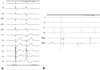
A 63-year-old man with symptomatic frequent PVCs was admitted for catheter ablation. His symptomatic PVCs were refractory to class I antiarrhythmic drugs for 1 year. The patient's medial history was unremarkable. Physical examination was normal except for an irregular heart rhythm. A 12-lead electrocardiogram (ECG) of PVC showed a monophasic R wave in leads 1 and aVL and precordial transition between leads V3 and V4 (Fig. 1A). Ambulatory ECG monitoring showed frequent PVC (55,882 beats; 30.1% of the total beats) and a type I second-degree atrioventricular (AV) block during sleep. Echocardiography showed normal biventricular size and function.
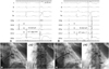
After informed consent was obtained from the patient, a cardiac electrophysiological study was performed under a fasting state. Sedation and analgesia were performed using midazolam and morphine. Baseline ECG showed sinus rhythm and PVC with the same configuration as clinical PVC. Programmed ventricular stimulation with or without isoproterenol did not induce sustained ventricular tachycardia. Activation mapping was performed using a 7-Fr conventional ablation catheter with a 4-mm distal tip. Local activation time at the His recording area was 18 ms ahead of PVC onset (Fig. 1B). Mapping of the right ventricle localized the PVC focus in the region of retroventricular (RV) inflow. The target site was initially mapped where the His potential had low amplitude and AV ratio is <1 to decrease the risk of a complete AV block. A site with local ventricular activation that preceded PVC onset by 24 ms was found anterior and inferior to the His recording catheter (Fig. 2A, B and C). The procedure was continued after we discussed the potential risk of a complete AV block with the patient.
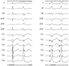
After radiofrequency (RF) energy was applied, a complete right bundle branch block occurred without affecting the PVC. Mapping of the His recording area more proximally revealed optimal mapping site with local ventricular activation ahead of PVC onset by 35 ms and the QS pattern on unipolar electrogram (Fig. 2D, E and F). Application of RF energy (escalated from 15 to 30 watts with a target temperature of 55℃) abolished the PVC within 4 seconds (Fig. 3). There was no junctional beat during the RF energy application. Pace mapping at the ablation site showed QRS configuration matches in all 12 leads (Fig. 4). PVC did not recur during the 60 minutes waiting period. Neither the PR nor the AH intervals changed before and after ablation. Total procedural time and fluoroscopic time was 180 and 20 minutes, respectively. The procedure was completed without further complications. Ambulatory monitoring showed a PVC burden of 0.3% and 0.0% at 1 month and 1 year of follow-up, respectively. A maximal heart rate of 140 beats per minute with normal AV conduction was noted during a treadmill exercise test 1 year after the procedure. The patient had no symptoms and did not require an antiarrhythmic drug during the 1-year follow-up period.
Discussion
Here we reported successful catheter ablation of a PVC from the proximal parahisian region without the creation of a complete AV block. Several investigators have developed algorithms to predict the origin of premature ventricular tachycardia and PVCs from the ventricular outflow tract using a 12-lead ECG.11)12) Based on this schema, the PVC origin in this patient is presumed to be located near the His bundle. Characteristics of PVC from the RV inflow tract include monophasic R waves in lead I, R, or RR in the aVL, the QS pattern in lead V1, and large R-wave amplitudes in leads V5-6.9)
Ashikaga et al.10) reported a case of parahisian PVC that was successfully treated by catheter ablation. Compared with previous reports, the configuration of PVC differed in the aspects of vertical PVC axis, deep S wave in lead III, a QRS pattern in leads V1 and V2, and a RR pattern in leads V4-6. This finding may be related to the PVC origin being from the more proximal His bundle region.
Successful ablation of PVC wherein the His potential is recorded from the right side suggests that the PVC originates from the myocardium in the vicinity of His at the right AV septum. Even though the AV ratio >1, absence of an AV block during and after ablation suggests that the PVC origin is very close to the distal penetrating bundle, which is surrounded by the central fibrous body. The central fibrous body may protect the His bundle from thermal injury caused by RF energy. Because the proximal penetrating bundle is not surrounded by the central fibrous body, ablation at this site may cause an AV block.13)
The existence of an AV block at night and the disappearance of the AV block during exercise suggests that the AV block is due to increased vagal tone. Normal AH and HV intervals also support the hypothesis that AV block is due to increased vagal tone. The idea that PVC induced the AV block by a mechanism of concealed conduction is less likely because there was no PVC at the time of the AV block and the second-degree AV block persisted after PVC ablation.
Catheter ablation in patients with parahisian PVC should be performed only after careful evaluation of PVC-related symptoms, a trial of maximally tolerated antiarrhythmic drug therapy, and full discussion about ablation-related catastrophic complications of the AV block. We performed the procedure because the patient's condition did not respond to standard antiarrhythmic drug treatment and he accepted the potential procedural risks.
Catheter ablation of arrhythmias originating in the vicinity of the bundle of His requires special attention to decrease the risk of iatrogenic AV block. In addition to careful mapping of the optimal target site, escalation of power from low energy, and immediate discontinuation of RF energy delivery immediately after the appearance of junctional rhythm are prerequisites to successful ablation.14) As in catheter ablation of the parahisian AV pathway, same precautions should be applied to avoid AV blocks during the ablation of parahisian PVC. Cryoablation can be considered a safer source of energy compared with RF energy in such patients at high risk of AV blocks.15)
There are several limitations in this study. We did not perform electroanatomic mapping, which might have revealed a ventricular substrate abnormality, or alternative site mapping from the left AV septum or right coronary cusp, which might have shown a more optimal site and had a lower risk of an AV block. We also did not map the anteroseptal region using the superior approach, which may have provided more stable and optimal target sites as did ablation of the anteroseptal accessory pathway.15)
In conclusion, we performed a successful catheter ablation of a PVC from the proximal His region under activation mapping guidance. Detailed mapping and careful delivery of RF energy are essential for successful ablation without AV block creation.
 XML Download