

 PDF
PDF ePub
ePub Citation
 Print
Print


Introduction
Although a lower incidence of major vascular access-related complications and the potential for early mobilization have led to increased use of transradial coronary angiography (TRA),1)2) the incidence of radial artery spasm (RAS), one of the most common complications of TRA, was reported up to 30%, and was associated with patient discomfort and lower procedural success rates.3-6) It has been reported that many factors are related to RAS, and the moderate-to-severe pain during radial artery cannulation is one of them.7) The radial artery is prone to catecholamine-induced contraction, because it has a predominance of α adrenoceptors.8) Therefore, any effort for reducing the radial pain during TRA could be beneficial for preventing RAS. A eutectic mixture of local anesthetic cream (EMLA®; Laboratoire ASTRA, Manterre, France), composed of lidocaine 2.5% and prilocaine 2.5%, is known to be an effective topical anesthetic agent. It is used for a variety of painful cutaneous procedures on intact skin, including phlebotomy, intravenous catheterization, arterial cannulation and lumbar puncture. Previously, Kim et al.9) have reported that, when applied 1 to 3 hours before the TRA, the EMLA cream can effectively reduce radial pain.
The primary objective of this study was to evaluate whether the EMLA cream, in addition to lidocaine infiltration, could reduce the sympathetic response by reducing radial pain during TRA. The secondary objective was to evaluate whether the EMLA cream could improve cannulation success rate and reduce the RAS in the study subjects.
Subjects and Methods
Study subjects
A total of 76 individuals, who were referred for coronary angiography from September 2008 to March 2009, were enrolled in this study. All patients were randomly assigned to either the EMLA group or the control (i.e., placebo cream) group, by a simple randomization table. All individuals and the physicians performing the coronary angiographies were blinded as to which cream was applied. This study was approved by the local Institutional Review Board (IRB approval number: CR308020) and all study participants signed informed consent forms.
Application of the study cream
We provided blinded tubes containing either EMLA cream or placebo, which was an odorless white cream that resembled the EMLA cream. 1 to 3 hours before TRA, either the EMLA 2.5 g (standard adult dose) or the placebo cream 2.5 g was applied to both wrists, 1 cm above the styloid process of the radius, and then covered with a transparent 5 cm dressing (Fig. 1).
Radial artery cannulation and coronary angiography
All procedures were performed by the operator experienced with TRA in 1,000 or more cases in one year.
After removing the applied cream, the radial artery was infiltrated with 0.6 mL of 2% lidocaine, using a 26-G needle, at a point located 5-10 mm proximal to the styloid process, and then punctured using a 20-G venous needle (Sindonbang Company, Korea). A 5-Fr 7 cm introducer sheath with hydrophilic coating (Terumo Company, Tokyo, Japan) was inserted into the radial artery. Heparin (5,000 IU) was administered into the radial artery via the introducer sheath. All patients underwent coronary angiography using 4-Fr Amplatz right (Jungsung Medical, Korea), 4-Fr Judkins or 5-Fr Judkins (MeritMedical, Ireland) angiographic catheters. The puncture site and the shape and size of the catheters were at operator's discretion.
Time and number of radial artery cannulations
We defined the cannulation time as the time from lidocaine infiltration to the puncture of the radial artery and counted the number of radial artery cannulations as the number of trials of radial artery puncture with the 26-G needle.
Estimating radial pain intensity
During lidocaine infiltration and introducer sheath insertion, pain intensity was assessed in each patient using a visual analogue scale (VAS) and a 4-category verbal rating scale (VRS-4).10) On the VAS, the patients indicated their pain intensity by making a mark on a 10-cm long line that included descriptors of pain intensity labeled at each end of the line (e.g., from "no pain" to "pain as bad as it could be"). The patient was instructed to regard the VAS as a continuum and to make a mark at the point along the line corresponding to his/her current level of pain. The score was determined by measuring the distance from the left end of the line to the patient's mark. Scoring of VRS-4 consists of a finite list of intensity descriptors: 1 point="no pain"; 2 points="a little pain"; 3 points="painful, but tolerable"; 4 points="most pain possible".
Measurement of the sympathetic response
Sympathetic tone, including systolic (SBP) and diastolic blood pressure (DBP), pulse rate (PR), stroke volume (SV) and total peripheral resistance (TPR), was measured continuously and non-invasively by finger photoplethysmography (Finometer®; Finapres Medical Systems, Netherlands). Previous studies showed that Finometer® recordings accurately reflect the sympathetic response.10-13)
We defined the sympathetic response as an absolute change of sympathetic tone at each step. The finger cuff of the Finometer® was applied to the mid-phalanx of the middle finger on the hand opposite to that with the vascular access site. To avoid hydrostatic level differences, the hand was continuously positioned at the right atrial level, in the mid-axillary line. After applying the Finometer®, the patient was stabilized for 5-10 minutes. We used as baseline values the mean values of sympathetic tone recorded for 1 minute before lidocaine infiltration. In the pilot phase of our study, we observed that the sympathetic tone started to increase 5-30 seconds after the noxious stimulus and decreased immediately after peaking. Because the time to peak differed among parameters, we used the peak value of each sympathetic parameter during the 30-40 seconds period after lidocaine infiltration, after arterial puncture and after introducer sheath insertion.
Angiographic definition of radial artery spasm
Retrograde radial angiography was performed three times: first, after insertion of the introducer sheath (i.e., at the start of procedure); second, after injection of 10 mL normal saline with nitroglycerine 200 µg (used as reference diameter); third, before withdrawal of the introducer sheath (i.e., at the end of procedure).
A 30 to 40-mm long segment from the tip of the introducer sheath at each step was selected to determine the diameter of the radial artery, using a computer-assisted quantification method (GE® Medical QCA, USA). We defined the reference diameter of the radial artery as the mean diameter of the radial artery after nitroglycerine injection. Angiographic RAS was defined as diameter stenosis ≥ 50% of the reference diameter at either the start or the end of procedure, using the following equations: diameter stenosis (DS, %) at the start of the procedure={reference diameter (RD)-minimal radial artery diameter (MD) after inserting sheath introducer}/RD×100 (%); DS (%) at the end of the procedure=(RD-MD before withdrawing sheath introducer)/RD×100 (%).
Clinical definition of radial artery spasm
The operator accessed the RAS on the basis of a questionnaire addressing the following four signs: persistent forearm pain, pain response to catheter manipulation, pain response to introducer withdrawal and difficult catheter manipulation after being trapped by the radial artery. Clinical RAS was defined as presence of at least 2 of these 4 signs or as the need, as determined by the operator, to administer a second dose of the spasmolytic agent.
Statistical analysis
Because there were no data for sympathetic response during TRA, we calculated the sample size for testing whether the EMLA cream reduces RAS during TRA based on a RAS incidence of 20% for the control group and 5% for the EMLA group, according to the results of previous studies.5)6) For inequality tests of the two proportions with a power of 80% and a two-sided α level of 0.05, we estimated that 36 subjects were needed in each group.
The statistical analysis was performed with the SPSS version 15 software (SPSS, Inc., Chicago, IL, USA). Continuous variables were expressed as mean±standard deviation, and categorical data as numbers (percentages). To compare values between the two groups, we used the Students t-test for continuous variables or the chi-square test for categorical variables.
Results
Baseline characteristics
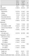
A total of 76 patients were enrolled in this study, 38 in each group. Patient baseline characteristics, including age, gender, medical history, medication, approach site, angiographic catheter used, anthropometric data, and reference diameter of the radial artery, were similar in the two groups. Baseline characteristics of study participants are presented in Table 1.
Radial pain score during the procedure
During lidocaine infiltration, the radial pain measured by VAS and VRS-4 was significantly lower in the EMLA group (VAS: 3.1±1.7 vs. 4.0±1.7, p=0.04; VRS-4: 2.0±0.5 vs. 2.2±0.5, p=0.03). However, during insertion of the introducer sheath, radial pain was similar in the two groups. Radial pain scores during the procedures are presented in Table 2.
Sympathetic response
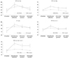
Sympathetic tone, including SBP, DBP, PR, SV and TPR measurements, were similar in the two groups at every step of the procedure. In contrast, from baseline to lidocaine infiltration, the sympathetic response was significantly blunted in the EMLA group (ΔSBP, mm Hg: 5 vs. 13, p<0.01; ΔDBP, mm Hg: 2 vs. 7, p=0.03; ΔPR, beat/min: 2 vs. 8, p<0.01, ΔSV, mL: 3 vs. 21, p<0.01; ΔTPR, mm Hg·L/min: 1.0 vs. 5.9, p<0.01); however, the difference, except for the change in SBP from lidocaine infiltration to puncture, was no longer present after lidocaine infiltration. Sympathetic responses from baseline to lidocaine infiltration, from lidocaine infiltration to puncture, and from puncture to sheath insertion, are presented in Table 3 and Fig. 2.
Radial artery cannulation and spasm
In the EMLA group, the cannulation time was significantly shorter (21.0±4.7 seconds. vs. 34.0±9.0 seconds., p<0.01) and the number of radial artery cannulations was significantly lower (1.21±0.47 vs. 1.50±0.69, p=0.04).
The rate of clinical or angiographic RAS was numerically lower in the EMLA group; however, the difference did not reach statistical significance. Although the rate of RAS was similar in the two groups, the minimal radial artery diameter (MD) at the start of procedure was significantly larger (1.78±0.54 mm vs. 1.51±0.41 mm, p=0.02) and the absolute change of diameter stenosis was significantly lower (11.9±8.4% vs. 18.3±12.7%, p=0.01) in the EMLA group. The results of radial artery cannulation and spasm are presented in Table 4.
Discussion
This study showed that the EMLA cream effectively reduces radial pain, and therefore blunts the sympathetic response during lidocaine infiltration; it also showed that the EMLA cream decreases the reduction of radial artery diameter at the start of procedure and therefore improves the cannulation success rate.
RAS is the most frequent complication of TRA and can cause significant discomfort to the patient, thereby reducing the procedural success rate.3)4) Even in centers with extensive experience with the radial approach, RAS can occur in 15 to 30% of TRA procedures.5)6) RAS may be related to radial artery anatomical anomalies, small diameter of the radial artery, prolonged cannulation, increased ratio of radial artery diameter to sheath outer diameter, anticoagulation treatment during arterial cannulation and moderate-to-severe pain during radial artery cannulation.7)14)
From the viewpoint of pain, radial artery is prone to spasm. Because the radial artery has a thick-walled vessel composed mainly of smooth muscle cells arranged in concentric layers and this marked muscular component of the artery, together with the high density of α1 receptors, makes this vessel especially susceptible to spasm.8) Therefore, the effort for reducing radial pain during procedure has an important role in preventing RAS. The radial pain reducing effect of the EMLA cream has been documented in several studies.9)15)16) Our study also shows significant reduction in radial pain, as measured by both VAS and VRS-4.
But none of the studies have evaluated the relationship between the pain and the sympathetic response. Our study uniquely evaluated the sympathetic response by measuring SBP, DBP, PR, SV, and TPR by non-invasive monitoring, and showed a significantly blunted sympathetic response in the EMLA group, during lidocaine infiltration. Certainly there is a limitation of our study because we did not measure the plasma levels of catecholamines, such as epinephrine and norepinephrine. However, these analyses are challenging, lack specificity and reproducibility, and present physiological limitations.17)
After lidocaine infiltration, however, we could not observe the difference in sympathetic response between the two groups. It is likely because lidocaine infiltration itself reduces radial pain and therefore the sympathetic response.18)
The role of adrenergic receptor is well known. The activation of α receptor results in peripheral vasoconstriction, and thus increases peripheral vascular resistance. Finally, these facts lead to increase DBP. In contrast, the activation of β receptors results in increased PR and SV, and thus increased cardiac output. Finally, these facts lead to increased SBP. In our study, we observed that all parameters related to α or β receptors were increasing after noxious stimuli. This finding reflects that the radial pain has a systemic effect on the patient, rather than a local effect on the radial artery.
Our study could not demonstrate a reduction in RAS by the EMLA cream. The reasons for this result could be explained as follows: First, because we used lidocaine infiltration in the two groups, the pain sensitivity would be equalized and this could result in no difference in RAS between two groups, especially after licocaine infiltration. Second, multiple factors other than pain, including number of radial artery punctures, type of catheter used, total procedure time, and patient's emotional status, might influence RAS incidence. Third, at the planning stage of this study, we calculated the sample size expecting 15% difference of RAS between the two groups. But the true incidence of RAS in our study was smaller than expected. So, if we increased the number of subjects, the statistical difference would be attained.
Although we could not demonstrate the reduction in RAS by the ELMA cream, we could observe the shortening of cannulation time and the reduction in the number of radial artery cannulations in this group. This could be achieved in the EMLA group by reducing the narrowing of the radial artery diameter at the start of the procedure.
In conclusion, the EMLA cream, in addition to lidocaine infiltration, effectively reduces the radial pain, and thus the sympathetic response, during lidocaine infiltration. With this benefit, the ELMA cream reduces the narrowing of the radial artery diameter at the start of procedure, and thus improves the cannulation in patients undergoing TRA.


 XML Download